3ZCJ

Crystal structure of Helicobacter pylori T4SS protein CagL in a tetragonal crystal form with a helical RGD-motif (6 Mol per ASU)


Experimental Data Snapshot

  • Method: X-RAY DIFFRACTION
  • Resolution: 3.25 Å
  • R-Value Free: 0.251 
  • R-Value Work: 0.205 
  • R-Value Observed: 0.207 

wwPDB Validation   3D Report Full Report


This is version 1.4 of the entry. See complete history


Literature

A Helical Rgd Motif Promoting Cell Adhesion: Crystal Structures of the Helicobacter Pylori Type Iv Secretion System Pilus Protein Cagl

Barden, S.Lange, S.Tegtmeyer, N.Conradi, J.Sewald, N.Backert, S.Niemann, H.H.

(2013) Structure 21: 1931

  • DOI: https://doi.org/10.1016/j.str.2013.08.018
  • Primary Citation of Related Structures:  
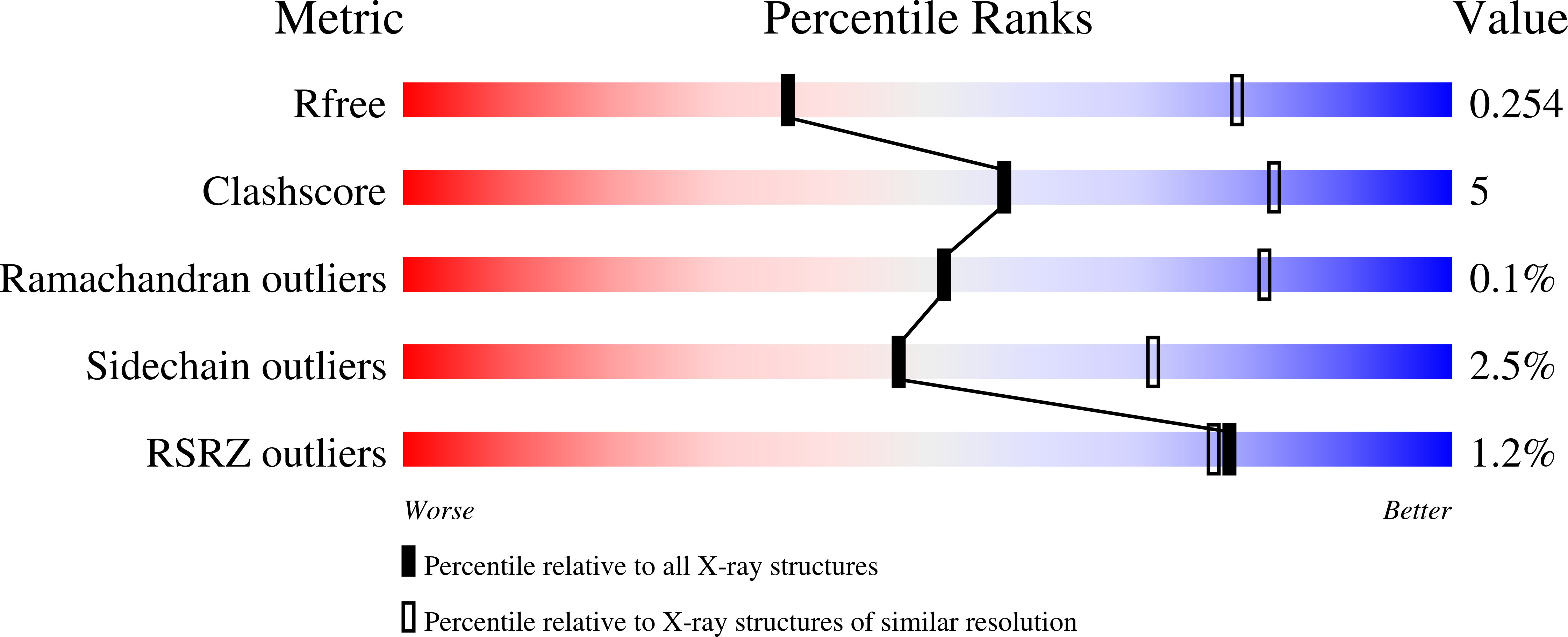
    3ZCI, 3ZCJ

  • PubMed Abstract: 

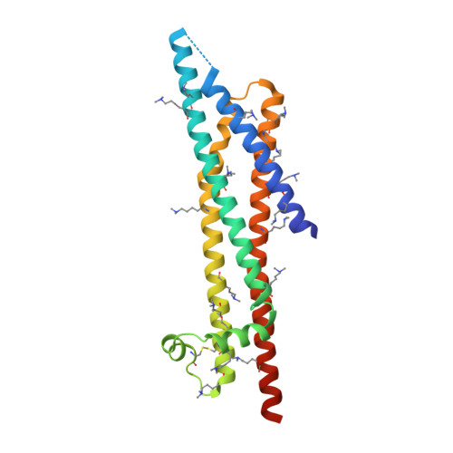
    RGD tripeptide motifs frequently mediate ligand binding to integrins. The type IV secretion system (T4SS) protein CagL of the gastric pathogen Helicobacter pylori also contains an RGD motif. CagL decorates the T4SS pilus and may function as an adhesin for host cells. Whether CagL binds integrins via its RGD motif is under debate. Here, we present crystal structures of CagL revealing an elongated four-helix bundle that appears evolutionarily unrelated to the proposed VirB5 orthologs. The RGD motif is surface-exposed but located within a long α helix. This is unprecedented as previously characterized integrin-binding RGD motifs are located within extended or flexible loops. Yet, adhesion of gastric epithelial cells to CagL was strictly RGD-dependent. Comparison of seven crystallographically independent molecules reveals substantial structural flexibility. Intramolecular disulfide bonds engineered to reduce CagL flexibility resulted in more stable protein, but unable to support cell adhesion. CagL may thus partly unfold during receptor binding.


  • Organizational Affiliation

    Structural Biochemistry, Department of Chemistry, Bielefeld University, 33501 Bielefeld, Germany.


Macromolecules
Find similar proteins by:  (by identity cutoff)  |  3D Structure
Entity ID: 1
MoleculeChains Sequence LengthOrganismDetailsImage
CAGL
A, B, C, D, E
A, B, C, D, E, F
220Helicobacter pylori 26695Mutation(s): 0 
UniProt
Find proteins for O25272 (Helicobacter pylori (strain ATCC 700392 / 26695))
Explore O25272 
Go to UniProtKB:  O25272
Entity Groups  
Sequence Clusters30% Identity50% Identity70% Identity90% Identity95% Identity100% Identity
UniProt GroupO25272
Sequence Annotations
Expand
  • Reference Sequence
Small Molecules
Ligands 4 Unique
IDChains Name / Formula / InChI Key2D Diagram3D Interactions
PEG
Query on PEG

Download Ideal Coordinates CCD File 
G [auth A]DI(HYDROXYETHYL)ETHER
C4 H10 O3
MTHSVFCYNBDYFN-UHFFFAOYSA-N
PO4
Query on PO4

Download Ideal Coordinates CCD File 
L [auth B],
R [auth F]
PHOSPHATE ION
O4 P
NBIIXXVUZAFLBC-UHFFFAOYSA-K
GOL
Query on GOL

Download Ideal Coordinates CCD File 
H [auth A]
I [auth A]
J [auth B]
K [auth B]
M [auth C]
H [auth A],
I [auth A],
J [auth B],
K [auth B],
M [auth C],
N [auth D],
P [auth E],
Q [auth F]
GLYCEROL
C3 H8 O3
PEDCQBHIVMGVHV-UHFFFAOYSA-N
K
Query on K

Download Ideal Coordinates CCD File 
O [auth D]POTASSIUM ION
K
NPYPAHLBTDXSSS-UHFFFAOYSA-N
Modified Residues  1 Unique
IDChains TypeFormula2D DiagramParent
MLY
Query on MLY
A, B, C, D, E
A, B, C, D, E, F
L-PEPTIDE LINKINGC8 H18 N2 O2LYS
Experimental Data & Validation

Experimental Data

  • Method: X-RAY DIFFRACTION
  • Resolution: 3.25 Å
  • R-Value Free: 0.251 
  • R-Value Work: 0.205 
  • R-Value Observed: 0.207 
  • Space Group: P 43 21 2
Unit Cell:
Length ( Å )Angle ( ˚ )
a = 197.122α = 90
b = 197.122β = 90
c = 106.808γ = 90
Software Package:
Software NamePurpose
PHENIXrefinement
XDSdata reduction
SCALAdata scaling
PHASERphasing

Structure Validation

View Full Validation Report


Entry History 

Deposition Data

Revision History  (Full details and data files)

  • Version 1.0: 2013-10-09
    Type: Initial release
  • Version 1.1: 2013-11-27
    Changes: Database references
  • Version 1.2: 2019-03-06
    Changes: Data collection, Derived calculations, Experimental preparation, Other
  • Version 1.3: 2019-09-25
    Changes: Data collection, Experimental preparation, Other
  • Version 1.4: 2023-12-20
    Changes: Data collection, Database references, Derived calculations, Refinement description