3VFD

Human spastin AAA domain


Experimental Data Snapshot

  • Method: X-RAY DIFFRACTION
  • Resolution: 3.30 Å
  • R-Value Free: 0.275 
  • R-Value Work: 0.206 
  • R-Value Observed: 0.213 

wwPDB Validation   3D Report Full Report


This is version 1.3 of the entry. See complete history


Literature

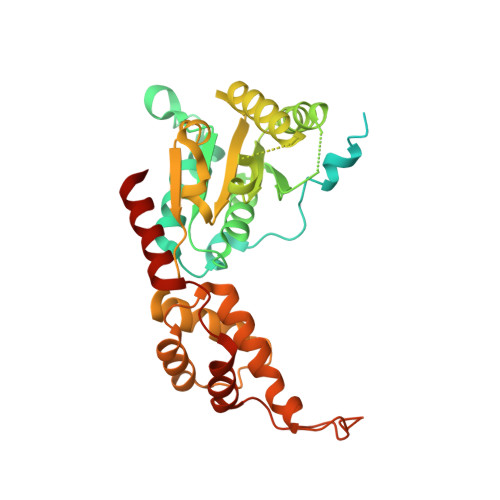
Crystal structure of the human spastin AAA domain.

Taylor, J.L.White, S.R.Lauring, B.Kull, F.J.

(2012) J Struct Biol 179: 133-137

  • DOI: https://doi.org/10.1016/j.jsb.2012.03.002
  • Primary Citation of Related Structures:  
    3VFD

  • PubMed Abstract: 

    Hereditary spastic paraplegia (HSP) is a motor neuron disease caused by a progressive degeneration of the motor axons of the corticospinal tract. Point mutations or exon deletions in the microtubule-severing ATPase, spastin, are responsible for approximately 40% of cases of autosomal dominant HSP. Here, we report the 3.3 Å X-ray crystal structure of a hydrolysis-deficient mutant (E442Q) of the human spastin protein AAA domain. This structure is analyzed in the context of the existing Drosophila melanogaster spastin AAA domain structure and crystal structures of other closely related proteins in order to build a more unifying framework for understanding the structural features of this group of microtubule-severing ATPases.


  • Organizational Affiliation

    Department of Chemistry, Dartmouth College, Hanover, NH 03755, USA.


Macromolecules
Find similar proteins by:  (by identity cutoff)  |  3D Structure
Entity ID: 1
MoleculeChains Sequence LengthOrganismDetailsImage
Spastin389Homo sapiensMutation(s): 1 
Gene Names: ADPSPFSP2KIAA1083SPASTSPG4
EC: 3.6.4.3
UniProt & NIH Common Fund Data Resources
Find proteins for Q9UBP0 (Homo sapiens)
Explore Q9UBP0 
Go to UniProtKB:  Q9UBP0
PHAROS:  Q9UBP0
GTEx:  ENSG00000021574 
Entity Groups  
Sequence Clusters30% Identity50% Identity70% Identity90% Identity95% Identity100% Identity
UniProt GroupQ9UBP0
Sequence Annotations
Expand
  • Reference Sequence
Small Molecules
Ligands 1 Unique
IDChains Name / Formula / InChI Key2D Diagram3D Interactions
SO4
Query on SO4

Download Ideal Coordinates CCD File 
B [auth A]SULFATE ION
O4 S
QAOWNCQODCNURD-UHFFFAOYSA-L
Experimental Data & Validation

Experimental Data

  • Method: X-RAY DIFFRACTION
  • Resolution: 3.30 Å
  • R-Value Free: 0.275 
  • R-Value Work: 0.206 
  • R-Value Observed: 0.213 
  • Space Group: P 65
Unit Cell:
Length ( Å )Angle ( ˚ )
a = 88.5α = 90
b = 88.5β = 90
c = 88.6γ = 120
Software Package:
Software NamePurpose
JBluIce-EPICSdata collection
PHENIXmodel building
PHENIXrefinement
XDSdata reduction
XSCALEdata scaling
PHENIXphasing

Structure Validation

View Full Validation Report


Entry History 

Deposition Data

Revision History  (Full details and data files)

  • Version 1.0: 2012-05-30
    Type: Initial release
  • Version 1.1: 2013-01-09
    Changes: Database references
  • Version 1.2: 2017-11-15
    Changes: Advisory, Refinement description
  • Version 1.3: 2023-09-13
    Changes: Advisory, Data collection, Database references, Derived calculations, Refinement description