


Structural adaptations of L-2-haloacid dehalogenases that enable hydrolytic defluorination
Chan, P.W.Y., To, T.K.W., Petit, P., Tran, C., Waelti, M., Savchenko, A., Yakunin, A.F., Edwards, E.A., Pai, E.F.To be published.
Experimental Data Snapshot
wwPDB Validation 3D Report Full Report

Entity ID: 1 | |||||
|---|---|---|---|---|---|
| Molecule | Chains | Sequence Length | Organism | Details | Image |
| Haloacid dehalogenase | 254 | Rhodococcus jostii RHA1 | Mutation(s): 0 Gene Names: RHA1_ro00230 EC: 3.8.1.2 | 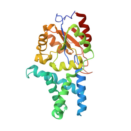 | |
UniProt | |||||
Find proteins for Q0SK70 (Rhodococcus jostii (strain RHA1)) Explore Q0SK70 Go to UniProtKB: Q0SK70 | |||||
Entity Groups | |||||
| Sequence Clusters | 30% Identity50% Identity70% Identity90% Identity95% Identity100% Identity | ||||
| UniProt Group | Q0SK70 | ||||
Sequence AnnotationsExpand | |||||
| |||||
| Ligands 1 Unique | |||||
|---|---|---|---|---|---|
| ID | Chains | Name / Formula / InChI Key | 2D Diagram | 3D Interactions | |
| CL Query on CL | I [auth A] J [auth C] K [auth D] L [auth E] M [auth F] | CHLORIDE ION Cl VEXZGXHMUGYJMC-UHFFFAOYSA-M |  | ||
| Length ( Å ) | Angle ( ˚ ) |
|---|---|
| a = 102.18 | α = 90 |
| b = 148.65 | β = 90 |
| c = 152.55 | γ = 90 |
| Software Name | Purpose |
|---|---|
| XSCALE | data scaling |
| REFMAC | refinement |
| PDB_EXTRACT | data extraction |
| XDS | data reduction |
| PHASER | phasing |
RCSB PDB (citation) is hosted by
RCSB PDB is a member of the

