


Structural adaptations of L-2-haloacid dehalogenases that enable hydrolytic defluorination
Chan, P.W.Y., To, T.K.W., Petit, P., Tran, C., Waelti, M., Savchenko, A., Yakunin, A.F., Edwards, E.A., Pai, E.F.To be published.
Experimental Data Snapshot
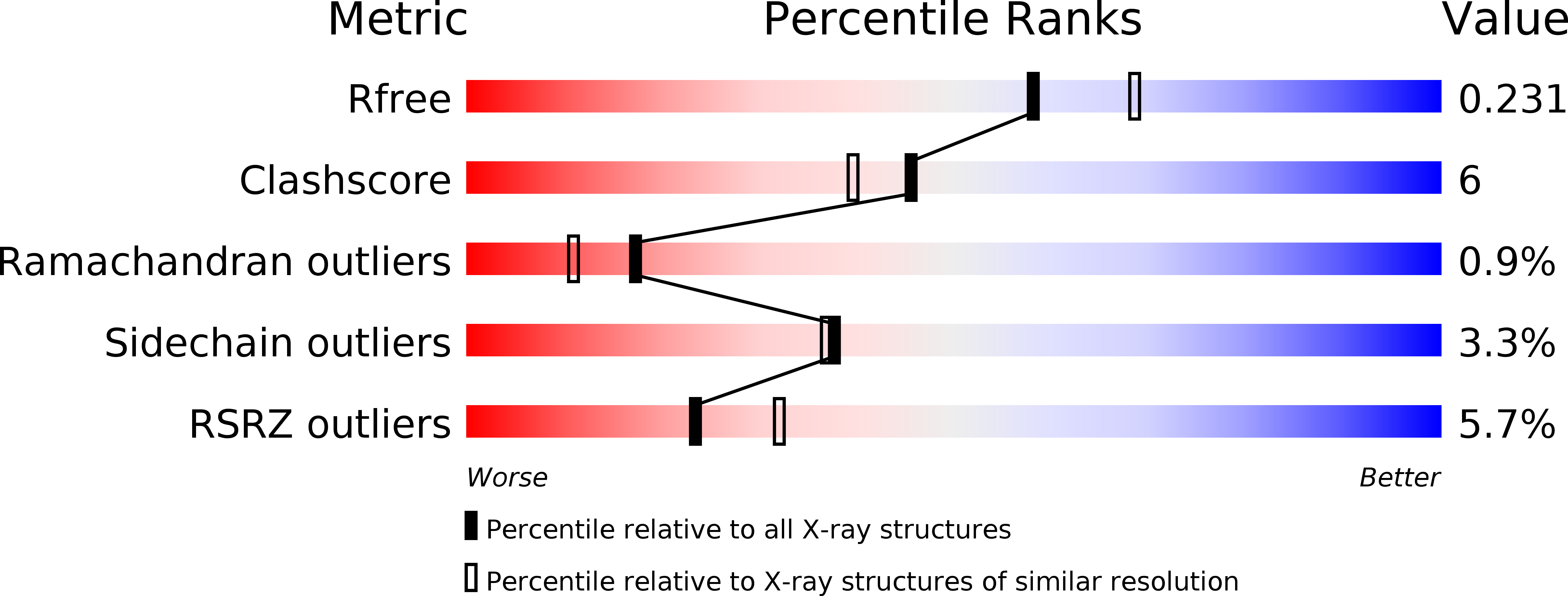
wwPDB Validation 3D Report Full Report
Entity ID: 1 | |||||
|---|---|---|---|---|---|
| Molecule | Chains | Sequence Length | Organism | Details | Image |
| haloacid dehalogenase | 254 | Pseudomonas aeruginosa | Mutation(s): 0 Gene Names: PA0810 EC: 3.8.1.2 | 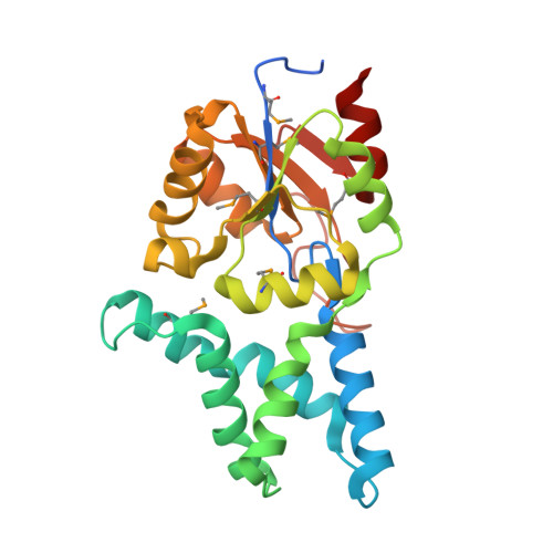 | |
UniProt | |||||
Find proteins for Q9I5C9 (Pseudomonas aeruginosa (strain ATCC 15692 / DSM 22644 / CIP 104116 / JCM 14847 / LMG 12228 / 1C / PRS 101 / PAO1)) Explore Q9I5C9 Go to UniProtKB: Q9I5C9 | |||||
Entity Groups | |||||
| Sequence Clusters | 30% Identity50% Identity70% Identity90% Identity95% Identity100% Identity | ||||
| UniProt Group | Q9I5C9 | ||||
Sequence AnnotationsExpand | |||||
| |||||
| Ligands 2 Unique | |||||
|---|---|---|---|---|---|
| ID | Chains | Name / Formula / InChI Key | 2D Diagram | 3D Interactions | |
| CL Query on CL | E [auth A], F [auth B], G [auth C], I [auth D] | CHLORIDE ION Cl VEXZGXHMUGYJMC-UHFFFAOYSA-M |  | ||
| NA Query on NA | H [auth C], J [auth D] | SODIUM ION Na FKNQFGJONOIPTF-UHFFFAOYSA-N |  | ||
| Modified Residues 1 Unique | |||||
|---|---|---|---|---|---|
| ID | Chains | Type | Formula | 2D Diagram | Parent |
| MSE Query on MSE | A, B, C, D | L-PEPTIDE LINKING | C5 H11 N O2 Se |  | MET |
| Length ( Å ) | Angle ( ˚ ) |
|---|---|
| a = 73.424 | α = 90 |
| b = 123.867 | β = 90.97 |
| c = 125.599 | γ = 90 |
| Software Name | Purpose |
|---|---|
| SCALA | data scaling |
| REFMAC | refinement |
| PDB_EXTRACT | data extraction |
| XDS | data reduction |
| SOLVE | phasing |
RCSB PDB (citation) is hosted by
RCSB PDB is a member of the

