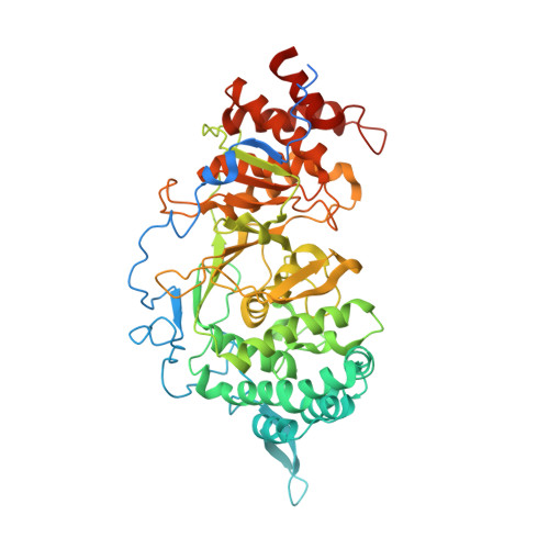
3UEL

Crystal structure of the catalytic domain of rat poly (ADP-ribose) glycohydrolase bound to ADP-HPD


Experimental Data Snapshot

  • Method: X-RAY DIFFRACTION
  • Resolution: 3.00 Å
  • R-Value Free: 0.274 
  • R-Value Work: 0.245 
  • R-Value Observed: 0.247 

wwPDB Validation   3D Report Full Report


Ligand Structure Quality Assessment 


This is version 1.2 of the entry. See complete history


Literature

Structure of mammalian poly(ADP-ribose) glycohydrolase reveals a flexible tyrosine clasp as a substrate-binding element.

Kim, I.K.Kiefer, J.R.Ho, C.M.Stegeman, R.A.Classen, S.Tainer, J.A.Ellenberger, T.

(2012) Nat Struct Mol Biol 19: 653-656

  • DOI: https://doi.org/10.1038/nsmb.2305
  • Primary Citation of Related Structures:  
    3UEK, 3UEL

  • PubMed Abstract: 

    Reversible post-translational modification by poly(ADP-ribose) (PAR) regulates chromatin structure, DNA repair and cell fate in response to genotoxic stress. PAR glycohydrolase (PARG) removes PAR chains from poly ADP-ribosylated proteins to restore protein function and release oligo(ADP-ribose) chains to signal damage. Here we report crystal structures of mammalian PARG and its complex with a substrate mimic that reveal an open substrate-binding site and a unique 'tyrosine clasp' enabling endoglycosidic cleavage of branched PAR chains.


  • Organizational Affiliation

    Department of Biochemistry and Molecular Biophysics, Washington University School of Medicine, St. Louis, Missouri, USA.


Macromolecules
Find similar proteins by:  (by identity cutoff)  |  3D Structure
Entity ID: 1
MoleculeChains Sequence LengthOrganismDetailsImage
Poly(ADP-ribose) glycohydrolase
A, B, C
588Rattus norvegicusMutation(s): 0 
Gene Names: Parg
EC: 3.2.1.143
UniProt
Find proteins for Q9QYM2 (Rattus norvegicus)
Explore Q9QYM2 
Go to UniProtKB:  Q9QYM2
Entity Groups  
Sequence Clusters30% Identity50% Identity70% Identity90% Identity95% Identity100% Identity
UniProt GroupQ9QYM2
Sequence Annotations
Expand
  • Reference Sequence
Small Molecules
Ligands 1 Unique
IDChains Name / Formula / InChI Key2D Diagram3D Interactions
A1R
Query on A1R

Download Ideal Coordinates CCD File 
D [auth A],
E [auth B],
F [auth C]
5'-O-[(S)-{[(S)-{[(2R,3R,4S)-3,4-DIHYDROXYPYRROLIDIN-2-YL]METHOXY}(HYDROXY)PHOSPHORYL]OXY}(HYDROXY)PHOSPHORYL]ADENOSINE
C15 H24 N6 O12 P2
NDQDTBCXPOIQGT-UHNJQBFTSA-N
Binding Affinity Annotations 
IDSourceBinding Affinity
A1R BindingDB:  3UEL IC50: min: 100, max: 1400 (nM) from 5 assay(s)
Experimental Data & Validation

Experimental Data

  • Method: X-RAY DIFFRACTION
  • Resolution: 3.00 Å
  • R-Value Free: 0.274 
  • R-Value Work: 0.245 
  • R-Value Observed: 0.247 
  • Space Group: C 2 2 21
Unit Cell:
Length ( Å )Angle ( ˚ )
a = 130.773α = 90
b = 195.989β = 90
c = 163.45γ = 90
Software Package:
Software NamePurpose
HKL-2000data collection
SOLVEphasing
REFMACrefinement
HKL-2000data reduction
SCALEPACKdata scaling

Structure Validation

View Full Validation Report


Ligand Structure Quality Assessment 


Entry History 

Deposition Data

Revision History  (Full details and data files)

  • Version 1.0: 2012-05-23
    Type: Initial release
  • Version 1.1: 2012-07-18
    Changes: Database references
  • Version 1.2: 2024-02-28
    Changes: Data collection, Database references, Derived calculations