3TBK

Mouse RIG-I ATPase Domain


Experimental Data Snapshot

  • Method: X-RAY DIFFRACTION
  • Resolution: 2.14 Å
  • R-Value Free: 0.236 
  • R-Value Work: 0.187 
  • R-Value Observed: 0.190 

wwPDB Validation   3D Report Full Report


Ligand Structure Quality Assessment 


This is version 1.2 of the entry. See complete history


Literature

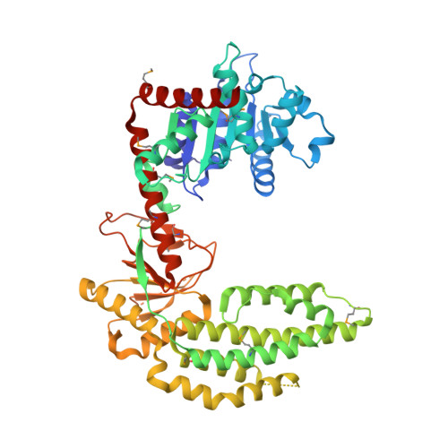
The RIG-I ATPase domain structure reveals insights into ATP-dependent antiviral signalling.

Civril, F.Bennett, M.Moldt, M.Deimling, T.Witte, G.Schiesser, S.Carell, T.Hopfner, K.P.

(2011) EMBO Rep 12: 1127-1134

  • DOI: https://doi.org/10.1038/embor.2011.190
  • Primary Citation of Related Structures:  
    3TBK

  • PubMed Abstract: 

    RIG-I detects cytosolic viral dsRNA with 5' triphosphates (5'-ppp-dsRNA), thereby initiating an antiviral innate immune response. Here we report the crystal structure of superfamily 2 (SF2) ATPase domain of RIG-I in complex with a nucleotide analogue. RIG-I SF2 comprises two RecA-like domains 1A and 2A and a helical insertion domain 2B, which together form a 'C'-shaped structure. Domains 1A and 2A are maintained in a 'signal-off' state with an inactive ATP hydrolysis site by an intriguing helical arm. By mutational analysis, we show surface motifs that are critical for dsRNA-stimulated ATPase activity, indicating that dsRNA induces a structural movement that brings domains 1A and 2A/B together to form an active ATPase site. The structure also indicates that the regulatory domain is close to the end of the helical arm, where it is well positioned to recruit 5'-ppp-dsRNA to the SF2 domain. Overall, our results indicate that the activation of RIG-I occurs through an RNA- and ATP-driven structural switch in the SF2 domain.


  • Organizational Affiliation

    Department of Biochemistry at the Gene Center, Ludwig-Maximilians-University Munich, Feodor-Lynen-Strasse 25, Munich 81377, Germany.


Macromolecules
Find similar proteins by:  (by identity cutoff)  |  3D Structure
Entity ID: 1
MoleculeChains Sequence LengthOrganismDetailsImage
RIG-I Helicase Domain555Mus musculusMutation(s): 0 
Gene Names: Ddx58
EC: 3.6.4.13
UniProt & NIH Common Fund Data Resources
Find proteins for Q6Q899 (Mus musculus)
Explore Q6Q899 
Go to UniProtKB:  Q6Q899
IMPC:  MGI:2442858
Entity Groups  
Sequence Clusters30% Identity50% Identity70% Identity90% Identity95% Identity100% Identity
UniProt GroupQ6Q899
Sequence Annotations
Expand
  • Reference Sequence
Small Molecules
Ligands 2 Unique
IDChains Name / Formula / InChI Key2D Diagram3D Interactions
ANP
Query on ANP

Download Ideal Coordinates CCD File 
B [auth A]PHOSPHOAMINOPHOSPHONIC ACID-ADENYLATE ESTER
C10 H17 N6 O12 P3
PVKSNHVPLWYQGJ-KQYNXXCUSA-N
EDO
Query on EDO

Download Ideal Coordinates CCD File 
C [auth A]1,2-ETHANEDIOL
C2 H6 O2
LYCAIKOWRPUZTN-UHFFFAOYSA-N
Modified Residues  1 Unique
IDChains TypeFormula2D DiagramParent
MSE
Query on MSE
A
L-PEPTIDE LINKINGC5 H11 N O2 SeMET
Experimental Data & Validation

Experimental Data

  • Method: X-RAY DIFFRACTION
  • Resolution: 2.14 Å
  • R-Value Free: 0.236 
  • R-Value Work: 0.187 
  • R-Value Observed: 0.190 
  • Space Group: P 21 21 21
Unit Cell:
Length ( Å )Angle ( ˚ )
a = 46.144α = 90
b = 86.027β = 90
c = 153.097γ = 90
Software Package:
Software NamePurpose
PHENIXmodel building
PHENIXrefinement
XDSdata reduction
XDSdata scaling
PHENIXphasing

Structure Validation

View Full Validation Report


Ligand Structure Quality Assessment 


Entry History 

Deposition Data

Revision History  (Full details and data files)

  • Version 1.0: 2011-10-26
    Type: Initial release
  • Version 1.1: 2011-12-14
    Changes: Database references
  • Version 1.2: 2017-11-08
    Changes: Refinement description