3RY7

Crystal Structure of Sa239


Experimental Data Snapshot

  • Method: X-RAY DIFFRACTION
  • Resolution: 2.15 Å
  • R-Value Free: 0.253 
  • R-Value Work: 0.216 
  • R-Value Observed: 0.218 

wwPDB Validation   3D Report Full Report


This is version 1.2 of the entry. See complete history


Literature

Crystal structure of Sa239 reveals the structural basis for the activation of ribokinase by monovalent cations.

Li, J.Wang, C.Wu, Y.Wu, M.Wang, L.Wang, Y.Zang, J.

(2012) J Struct Biol 177: 578-582

  • DOI: https://doi.org/10.1016/j.jsb.2011.12.010
  • Primary Citation of Related Structures:  
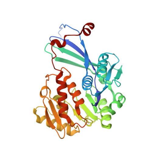
    3RY7

  • PubMed Abstract: 

    Ribokinase is responsible for catalyzing the reaction of d-ribose and ATP to produce ribose-5-phosphate and ADP, which can be activated by monovalent cations such as potassium, cesium and ammonium. However, the exact activation mechanism of ribokinase remains elusive. Here we report the crystal structure of Sa239, a ribokinase from Staphylococcus aureus, in the absence of monovalent ions. In addition to the dimer form similar to that observed in Escherichia coli ribokinase structure, the structure of Sa239 demonstrates that the C-terminal tail protrudes from the remaining part and interacts with the neighboring molecule, resulting in an unexpected dimerization form. By comparing the structure of Sa239 to E. coli ribokinase, we propose that binding of the monovalent cation triggers the conformational change of the large ATP loop to organize the formation of nucleotide binding pocket, thus enabling ATP binding and enhancing catalytic activity. Our study uncovers the detailed structural basis for the activation mechanism of ribokinase by monovalent cations.


  • Organizational Affiliation

    Hefei National Laboratory for Physical Sciences at Microscale, School of Life Sciences, University of Science and Technology of China, 96 Jinzhai Road, Hefei, Anhui 230026, People's Republic of China.


Macromolecules
Find similar proteins by:  (by identity cutoff)  |  3D Structure
Entity ID: 1
MoleculeChains Sequence LengthOrganismDetailsImage
Ribokinase304Staphylococcus aureus subsp. aureus COLMutation(s): 0 
Gene Names: rbsKSACOL0253
EC: 2.7.1.15
UniProt
Find proteins for A0A0H2WZY4 (Staphylococcus aureus (strain COL))
Explore A0A0H2WZY4 
Go to UniProtKB:  A0A0H2WZY4
Entity Groups  
Sequence Clusters30% Identity50% Identity70% Identity90% Identity95% Identity100% Identity
UniProt GroupA0A0H2WZY4
Sequence Annotations
Expand
  • Reference Sequence
Experimental Data & Validation

Experimental Data

  • Method: X-RAY DIFFRACTION
  • Resolution: 2.15 Å
  • R-Value Free: 0.253 
  • R-Value Work: 0.216 
  • R-Value Observed: 0.218 
  • Space Group: P 65 2 2
Unit Cell:
Length ( Å )Angle ( ˚ )
a = 93.752α = 90
b = 93.752β = 90
c = 165.27γ = 120
Software Package:
Software NamePurpose
MAR345dtbdata collection
PHASERphasing
REFMACrefinement
HKL-2000data reduction
HKL-2000data scaling

Structure Validation

View Full Validation Report


Entry History 

Deposition Data

Revision History  (Full details and data files)

  • Version 1.0: 2012-04-25
    Type: Initial release
  • Version 1.1: 2016-12-21
    Changes: Structure summary
  • Version 1.2: 2023-11-01
    Changes: Data collection, Database references, Derived calculations, Refinement description