3P3E

Crystal Structure of the PSEUDOMONAS AERUGINOSA LpxC/LPC-009 complex


Experimental Data Snapshot

  • Method: X-RAY DIFFRACTION
  • Resolution: 1.28 Å
  • R-Value Free: 0.193 
  • R-Value Work: 0.182 
  • R-Value Observed: 0.182 

wwPDB Validation   3D Report Full Report


Ligand Structure Quality Assessment 


This is version 1.4 of the entry. See complete history


Literature

Species-specific and inhibitor-dependent conformations of LpxC: implications for antibiotic design.

Lee, C.J.Liang, X.Chen, X.Zeng, D.Joo, S.H.Chung, H.S.Barb, A.W.Swanson, S.M.Nicholas, R.A.Li, Y.Toone, E.J.Raetz, C.R.Zhou, P.

(2011) Chem Biol 18: 38-47

  • DOI: https://doi.org/10.1016/j.chembiol.2010.11.011
  • Primary Citation of Related Structures:  
    3P3C, 3P3E, 3P3G

  • PubMed Abstract: 

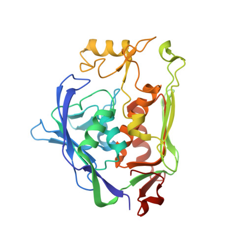
    LpxC is an essential enzyme in the lipid A biosynthetic pathway in gram-negative bacteria. Several promising antimicrobial lead compounds targeting LpxC have been reported, though they typically display a large variation in potency against different gram-negative pathogens. We report that inhibitors with a diacetylene scaffold effectively overcome the resistance caused by sequence variation in the LpxC substrate-binding passage. Compound binding is captured in complex with representative LpxC orthologs, and structural analysis reveals large conformational differences that mostly reflect inherent molecular features of distinct LpxC orthologs, whereas ligand-induced structural adaptations occur at a smaller scale. These observations highlight the need for a molecular understanding of inherent structural features and conformational plasticity of LpxC enzymes for optimizing LpxC inhibitors as broad-spectrum antibiotics against gram-negative infections.


  • Organizational Affiliation

    Department of Biochemistry, Duke University Medical Center, Durham, NC 27710, USA.


Macromolecules
Find similar proteins by:  (by identity cutoff)  |  3D Structure
Entity ID: 1
MoleculeChains Sequence LengthOrganismDetailsImage
UDP-3-O-[3-hydroxymyristoyl] N-acetylglucosamine deacetylase299Pseudomonas aeruginosaMutation(s): 1 
Gene Names: envAlpxCPA4406
EC: 3.5.1
UniProt
Find proteins for P47205 (Pseudomonas aeruginosa (strain ATCC 15692 / DSM 22644 / CIP 104116 / JCM 14847 / LMG 12228 / 1C / PRS 101 / PAO1))
Explore P47205 
Go to UniProtKB:  P47205
Entity Groups  
Sequence Clusters30% Identity50% Identity70% Identity90% Identity95% Identity100% Identity
UniProt GroupP47205
Sequence Annotations
Expand
  • Reference Sequence
Small Molecules
Ligands 4 Unique
IDChains Name / Formula / InChI Key2D Diagram3D Interactions
3P3
Query on 3P3

Download Ideal Coordinates CCD File 
D [auth A]N-[(1S,2R)-2-hydroxy-1-(hydroxycarbamoyl)propyl]-4-(4-phenylbuta-1,3-diyn-1-yl)benzamide
C21 H18 N2 O4
VUYMSCCEGRLBAF-BEFAXECRSA-N
ZN
Query on ZN

Download Ideal Coordinates CCD File 
B [auth A],
C [auth A]
ZINC ION
Zn
PTFCDOFLOPIGGS-UHFFFAOYSA-N
NO3
Query on NO3

Download Ideal Coordinates CCD File 
AA [auth A]
BA [auth A]
CA [auth A]
DA [auth A]
F [auth A]
AA [auth A],
BA [auth A],
CA [auth A],
DA [auth A],
F [auth A],
G [auth A],
H [auth A],
I [auth A],
J [auth A],
K [auth A],
L [auth A],
M [auth A],
N [auth A],
O [auth A],
P [auth A],
Q [auth A],
R [auth A],
S [auth A],
T [auth A],
U [auth A],
V [auth A],
W [auth A],
X [auth A],
Y [auth A],
Z [auth A]
NITRATE ION
N O3
NHNBFGGVMKEFGY-UHFFFAOYSA-N
NA
Query on NA

Download Ideal Coordinates CCD File 
E [auth A]SODIUM ION
Na
FKNQFGJONOIPTF-UHFFFAOYSA-N
Binding Affinity Annotations 
IDSourceBinding Affinity
3P3 BindingDB:  3P3E Kd: 8.35 (nM) from 1 assay(s)
Experimental Data & Validation

Experimental Data

  • Method: X-RAY DIFFRACTION
  • Resolution: 1.28 Å
  • R-Value Free: 0.193 
  • R-Value Work: 0.182 
  • R-Value Observed: 0.182 
  • Space Group: P 21 21 21
Unit Cell:
Length ( Å )Angle ( ˚ )
a = 52.637α = 90
b = 73.608β = 90
c = 88.308γ = 90
Software Package:
Software NamePurpose
DENZOdata reduction
SCALEPACKdata scaling
PHASERphasing
PHENIXrefinement
PDB_EXTRACTdata extraction

Structure Validation

View Full Validation Report


Ligand Structure Quality Assessment 


Entry History 

Deposition Data

Revision History  (Full details and data files)

  • Version 1.0: 2011-01-05
    Type: Initial release
  • Version 1.1: 2011-07-13
    Changes: Version format compliance
  • Version 1.2: 2017-11-08
    Changes: Advisory, Refinement description
  • Version 1.3: 2018-01-24
    Changes: Structure summary
  • Version 1.4: 2024-02-21
    Changes: Advisory, Data collection, Database references, Derived calculations