3M9E

Thyroid hormone beta DNA binding domain homodimer with inverted palindrome TRE


Experimental Data Snapshot

  • Method: X-RAY DIFFRACTION
  • Resolution: 2.41 Å
  • R-Value Free: 0.231 
  • R-Value Work: 0.179 
  • R-Value Observed: 0.182 

wwPDB Validation   3D Report Full Report


This is version 1.2 of the entry. See complete history


Literature

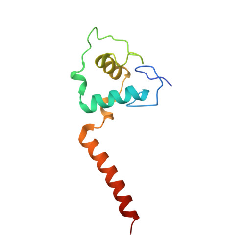
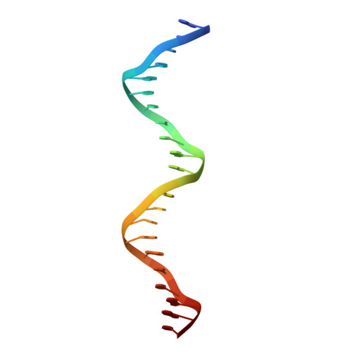
Structure of a thyroid hormone receptor DNA-binding domain homodimer bound to an inverted palindrome DNA response element.

Chen, Y.Young, M.A.

(2010) Mol Endocrinol 24: 1650-1664

  • DOI: https://doi.org/10.1210/me.2010-0129
  • Primary Citation of Related Structures:  
    3M9E

  • PubMed Abstract: 

    Thyroid hormone receptor (TR), as a member of the nuclear hormone receptor family, can recognize and bind different classes of DNA response element targets as either a monomer, a homooligomer, or a heterooligomer. We report here the first crystal structure of a homodimer TR DNA-binding domain (DBD) in complex with an inverted repeat class of thyroid response element (TRE). The structure shows a nearly symmetric structure of the TR DBD assembled on the F2 TRE where the base recognition contacts in the homodimer DNA complex are conserved relative to the previously published structure of a TR-9-cis-retinoic acid receptor heterodimer DNA complex. The new structure also reveals that the T-box region of the DBD can function as a structural hinge that enables a large degree of flexibility in the position of the C-terminal extension helix that connects the DBD to the ligand-binding domain. Although the isolated TR DBDs exist as monomers in solution, we have measured highly cooperative binding of the two TR DBD subunits onto the inverted repeat DNA sequence. This suggests that elements of the DBD can influence the specific TR oligomerization at target genes, and it is not just interactions between the ligand-binding domains that are responsible for TR oligomerization at target genes. Mutational analysis shows that intersubunit contacts at the DBD C terminus account for some, but not all, of the cooperative homodimer TR binding to the inverted repeat class TRE.


  • Organizational Affiliation

    Department of Biophysics, University of Michigan, Ann Arbor, Michigan 48109, USA.


Macromolecules

Find similar proteins by:  (by identity cutoff)  |  3D Structure
Entity ID: 1
MoleculeChains Sequence LengthOrganismDetailsImage
Thyroid hormone receptor beta
A, B, E, F
105Rattus norvegicusMutation(s): 0 
Gene Names: Erba2Nr1a2RODThrb
UniProt
Find proteins for P18113 (Rattus norvegicus)
Explore P18113 
Go to UniProtKB:  P18113
Entity Groups  
Sequence Clusters30% Identity50% Identity70% Identity90% Identity95% Identity100% Identity
UniProt GroupP18113
Sequence Annotations
Expand
  • Reference Sequence

Find similar nucleic acids by:  Sequence   |   3D Structure  

Entity ID: 2
MoleculeChains LengthOrganismImage
DNA (5'-D(*AP*TP*TP*GP*AP*CP*CP*TP*CP*AP*GP*CP*TP*GP*AP*GP*GP*TP*CP*AP*AP*T)-3')
C, D, G, H
22N/A
Sequence Annotations
Expand
  • Reference Sequence
Experimental Data & Validation

Experimental Data

  • Method: X-RAY DIFFRACTION
  • Resolution: 2.41 Å
  • R-Value Free: 0.231 
  • R-Value Work: 0.179 
  • R-Value Observed: 0.182 
  • Space Group: P 1 21 1
Unit Cell:
Length ( Å )Angle ( ˚ )
a = 73.738α = 90
b = 83.468β = 98.28
c = 75.743γ = 90
Software Package:
Software NamePurpose
HKL-2000data collection
PHENIXmodel building
PHENIXrefinement
HKL-2000data reduction
HKL-2000data scaling
PHENIXphasing

Structure Validation

View Full Validation Report


Entry History 

Deposition Data

  • Released Date: 2010-08-18 
  • Deposition Author(s): Chen, Y.

Revision History  (Full details and data files)

  • Version 1.0: 2010-08-18
    Type: Initial release
  • Version 1.1: 2011-07-13
    Changes: Version format compliance
  • Version 1.2: 2023-09-06
    Changes: Data collection, Database references, Derived calculations, Refinement description