


Crystal structure of transketolase in complex with thiamine diphosphate, ribose-5-phosphate and calcium ion
Nocek, B., Makowska-Grzyska, M., Maltseva, N., Grimshaw, S., Joachimiak, A., Anderson, W.To be published.
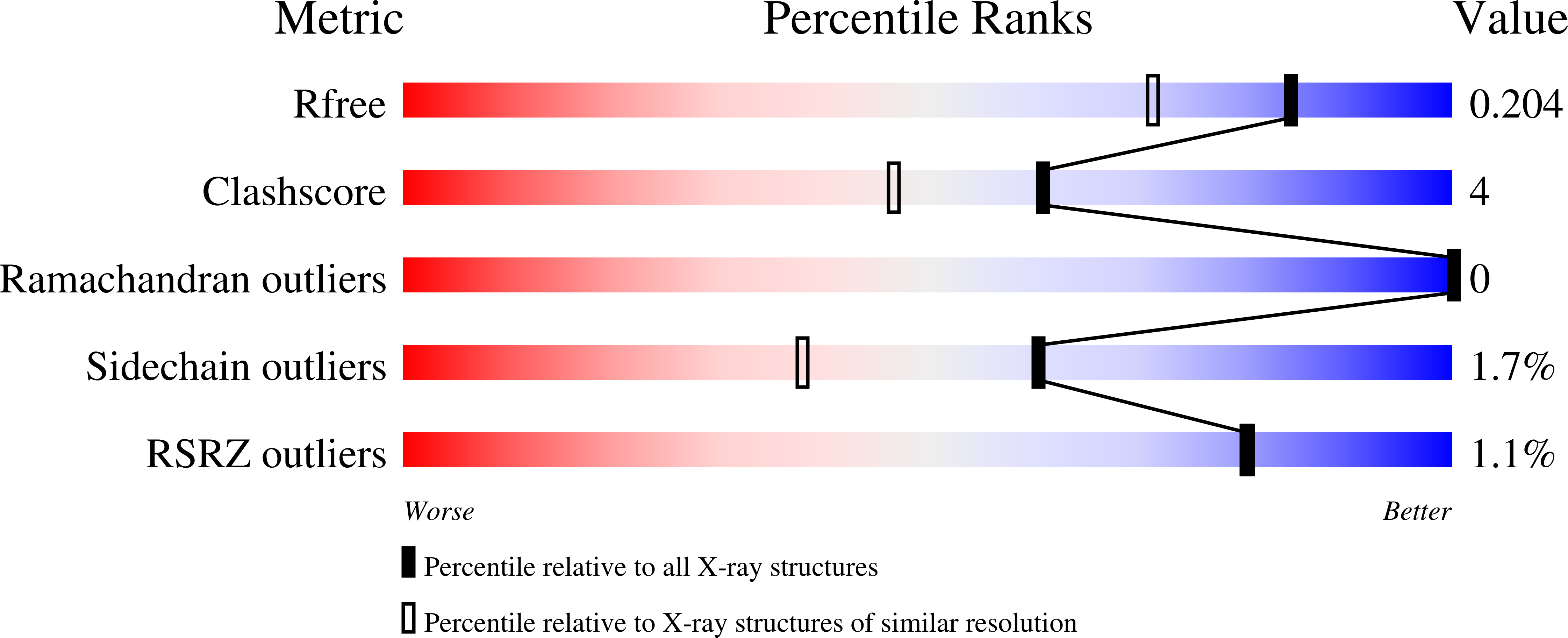
Experimental Data Snapshot
Entity ID: 1 | |||||
|---|---|---|---|---|---|
| Molecule | Chains | Sequence Length | Organism | Details | Image |
| Transketolase | 635 | Campylobacter jejuni subsp. jejuni NCTC 11168 = ATCC 700819 | Mutation(s): 0 Gene Names: tkt, Cj1645 EC: 2.2.1.1 | 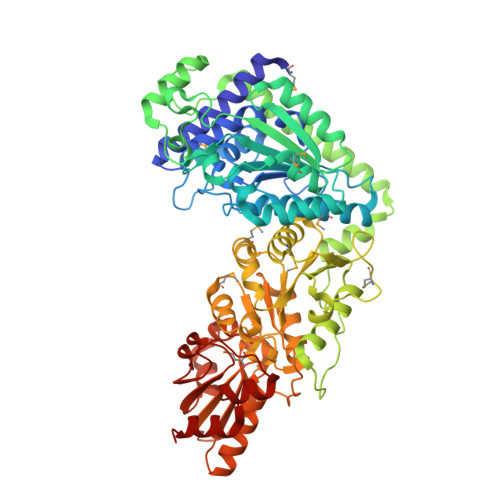 | |
UniProt | |||||
Find proteins for Q0P7Y3 (Campylobacter jejuni subsp. jejuni serotype O:2 (strain ATCC 700819 / NCTC 11168)) Explore Q0P7Y3 Go to UniProtKB: Q0P7Y3 | |||||
Entity Groups | |||||
| Sequence Clusters | 30% Identity50% Identity70% Identity90% Identity95% Identity100% Identity | ||||
| UniProt Group | Q0P7Y3 | ||||
Sequence AnnotationsExpand | |||||
| |||||
| Ligands 6 Unique | |||||
|---|---|---|---|---|---|
| ID | Chains | Name / Formula / InChI Key | 2D Diagram | 3D Interactions | |
| TPP Query on TPP | C [auth A] | THIAMINE DIPHOSPHATE C12 H19 N4 O7 P2 S AYEKOFBPNLCAJY-UHFFFAOYSA-O |  | ||
| R5P Query on R5P | F [auth A] | RIBOSE-5-PHOSPHATE C5 H11 O8 P PPQRONHOSHZGFQ-LMVFSUKVSA-N |  | ||
| PO4 Query on PO4 | G [auth A] | PHOSPHATE ION O4 P NBIIXXVUZAFLBC-UHFFFAOYSA-K |  | ||
| GOL Query on GOL | B [auth A] | GLYCEROL C3 H8 O3 PEDCQBHIVMGVHV-UHFFFAOYSA-N |  | ||
| EDO Query on EDO | E [auth A] | 1,2-ETHANEDIOL C2 H6 O2 LYCAIKOWRPUZTN-UHFFFAOYSA-N |  | ||
| CA Query on CA | D [auth A] | CALCIUM ION Ca BHPQYMZQTOCNFJ-UHFFFAOYSA-N |  | ||
| Modified Residues 1 Unique | |||||
|---|---|---|---|---|---|
| ID | Chains | Type | Formula | 2D Diagram | Parent |
| MSE Query on MSE | A | L-PEPTIDE LINKING | C5 H11 N O2 Se |  | MET |
| Length ( Å ) | Angle ( ˚ ) |
|---|---|
| a = 130.924 | α = 90 |
| b = 71.462 | β = 109.48 |
| c = 69.617 | γ = 90 |
| Software Name | Purpose |
|---|---|
| SBC-Collect | data collection |
| MOLREP | phasing |
| Coot | model building |
| REFMAC | refinement |
| HKL-3000 | data reduction |
| HKL-3000 | data scaling |
RCSB PDB (citation) is hosted by
RCSB PDB is a member of the

