3M2M

Rat galectin-1 complex with lactose


Experimental Data Snapshot

  • Method: X-RAY DIFFRACTION
  • Resolution: 2.95 Å
  • R-Value Free: 0.248 
  • R-Value Work: 0.206 
  • R-Value Observed: 0.233 

wwPDB Validation   3D Report Full Report


This is version 2.0 of the entry. See complete history


Literature

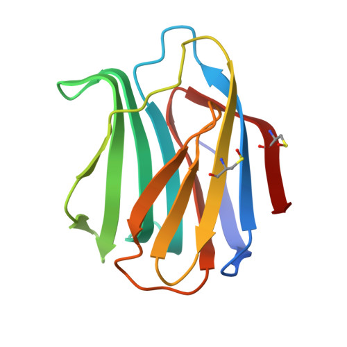
Monovalent interactions of galectin-1.

Salomonsson, E.Larumbe, A.Tejler, J.Tullberg, E.Rydberg, H.Sundin, A.Khabut, A.Frejd, T.Lobsanov, Y.D.Rini, J.M.Nilsson, U.J.Leffler, H.

(2010) Biochemistry 49: 9518-9532

  • DOI: https://doi.org/10.1021/bi1009584
  • Primary Citation of Related Structures:  
    3M2M

  • PubMed Abstract: 

    Galectin-1, a β-galactoside binding lectin involved in immunoregulation and cancer, binds natural and many synthetic multivalent glycoconjugates with an apparent glycoside cluster effect, that is, affinity above and beyond what would be expected from the concentration of the determinant sugar. Here we have analyzed the mechanism of such cluster effects in solution at physiological concentration using a fluorescence anisotropy assay with a novel fluorescent high-affinity galectin-1 binding probe. The interaction of native dimeric and monomeric mutants of rat and human galectin-1 with mono- and divalent small molecules, fetuin, asialofetuin, and human serum glycoproteins was analyzed. Surprisingly, high-affinity binding did not depend much on the dimeric state of galectin-1 and thus is due mainly to monomeric interactions of a single carbohydrate recognition domain. The mechanism for this is unknown, but one possibility includes additional interactions that high-affinity ligands make with an extended binding site on the carbohydrate recognition domain. It follows that such weak additional interactions must be important for the biological function of galectin-1 and also for the design of galectin-1 inhibitors.


  • Organizational Affiliation

    Institute of Laboratory Medicine, Lund University, Lund, Sweden.


Macromolecules
Find similar proteins by:  (by identity cutoff)  |  3D Structure
Entity ID: 1
MoleculeChains Sequence LengthOrganismDetailsImage
Galectin-1
A, B, C, D, E
A, B, C, D, E, F, G, H
134Rattus norvegicusMutation(s): 0 
Gene Names: Lgals1
UniProt
Find proteins for P11762 (Rattus norvegicus)
Explore P11762 
Go to UniProtKB:  P11762
Entity Groups  
Sequence Clusters30% Identity50% Identity70% Identity90% Identity95% Identity100% Identity
UniProt GroupP11762
Sequence Annotations
Expand
  • Reference Sequence
Oligosaccharides

Help

Entity ID: 2
MoleculeChains Length2D Diagram Glycosylation3D Interactions
beta-D-galactopyranose-(1-4)-beta-D-glucopyranose
I, J, K, L, M
2N/A
Glycosylation Resources
GlyTouCan:  G84224TW
GlyCosmos:  G84224TW
GlyGen:  G84224TW
Small Molecules
Modified Residues  1 Unique
IDChains TypeFormula2D DiagramParent
CSO
Query on CSO
A, B, C, D, E
A, B, C, D, E, F, G, H
L-PEPTIDE LINKINGC3 H7 N O3 SCYS
Experimental Data & Validation

Experimental Data

  • Method: X-RAY DIFFRACTION
  • Resolution: 2.95 Å
  • R-Value Free: 0.248 
  • R-Value Work: 0.206 
  • R-Value Observed: 0.233 
  • Space Group: P 1
Unit Cell:
Length ( Å )Angle ( ˚ )
a = 40.1α = 101.1
b = 58.6β = 91.6
c = 121.6γ = 110.9
Software Package:
Software NamePurpose
CNSrefinement
PDB_EXTRACTdata extraction
XDSdata scaling
XDSdata reduction
XSCALEdata scaling
MERLOTphasing
GLRFphasing
INTREFphasing
X-PLORphasing
AMoREphasing

Structure Validation

View Full Validation Report


Entry History 

Deposition Data

Revision History  (Full details and data files)

  • Version 1.0: 2010-07-21
    Type: Initial release
  • Version 1.1: 2011-07-13
    Changes: Version format compliance
  • Version 2.0: 2020-07-29
    Type: Remediation
    Reason: Carbohydrate remediation
    Changes: Atomic model, Data collection, Derived calculations, Non-polymer description, Structure summary