


Crystal structure of glycosyl hydrolase family 5 (NP_809925.1) from BACTEROIDES THETAIOTAOMICRON VPI-5482 at 2.10 A resolution
Joint Center for Structural Genomics (JCSG)To be published.
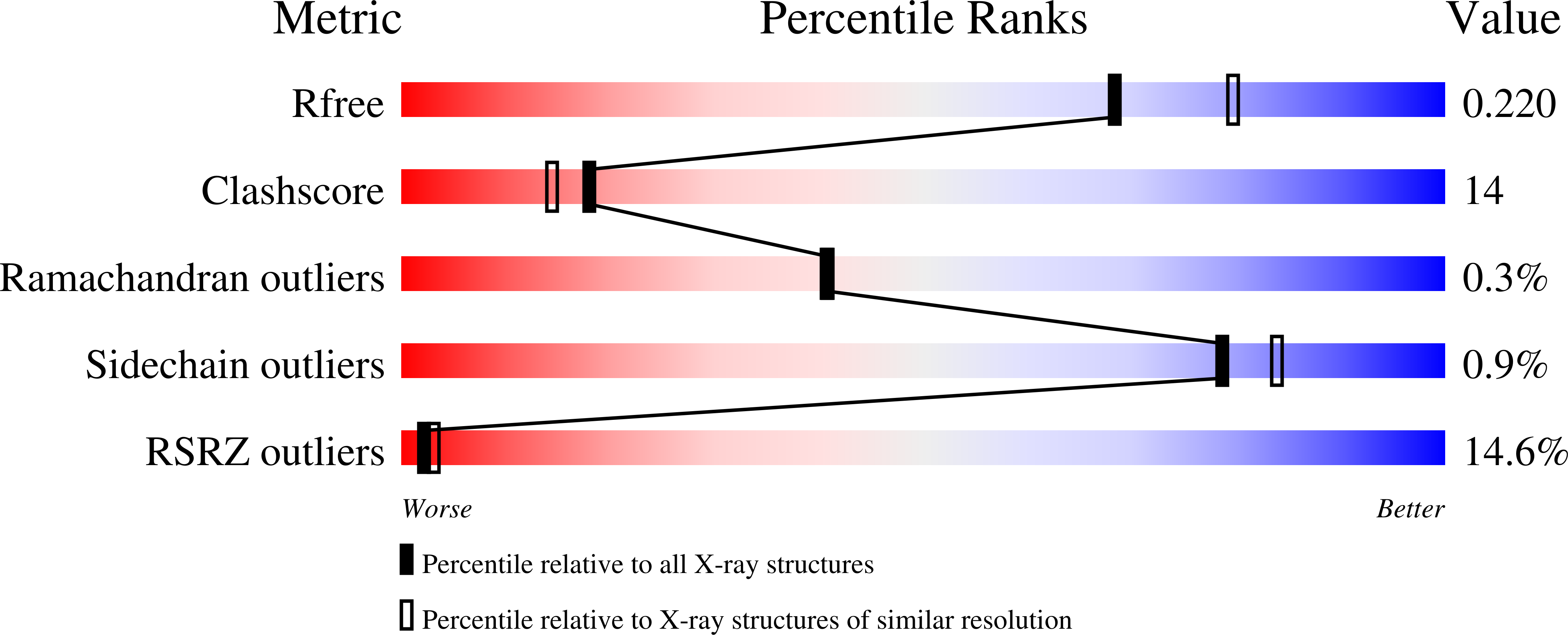
Experimental Data Snapshot
wwPDB Validation 3D Report Full Report
Entity ID: 1 | |||||
|---|---|---|---|---|---|
| Molecule | Chains | Sequence Length | Organism | Details | Image |
| glycosyl hydrolase family 5 | 463 | Bacteroides thetaiotaomicron VPI-5482 | Mutation(s): 0 Gene Names: BT_1012 | 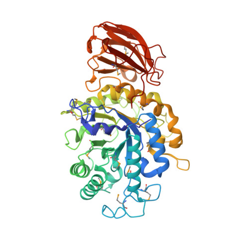 | |
UniProt | |||||
Find proteins for Q8A905 (Bacteroides thetaiotaomicron (strain ATCC 29148 / DSM 2079 / JCM 5827 / CCUG 10774 / NCTC 10582 / VPI-5482 / E50)) Explore Q8A905 Go to UniProtKB: Q8A905 | |||||
Entity Groups | |||||
| Sequence Clusters | 30% Identity50% Identity70% Identity90% Identity95% Identity100% Identity | ||||
| UniProt Group | Q8A905 | ||||
Sequence AnnotationsExpand | |||||
| |||||
| Ligands 3 Unique | |||||
|---|---|---|---|---|---|
| ID | Chains | Name / Formula / InChI Key | 2D Diagram | 3D Interactions | |
| MPD Query on MPD | AA [auth C], DA [auth C] | (4S)-2-METHYL-2,4-PENTANEDIOL C6 H14 O2 SVTBMSDMJJWYQN-YFKPBYRVSA-N |  | ||
| MRD Query on MRD | BA [auth C] CA [auth C] K [auth A] L [auth A] M [auth A] | (4R)-2-METHYLPENTANE-2,4-DIOL C6 H14 O2 SVTBMSDMJJWYQN-RXMQYKEDSA-N |  | ||
| SO4 Query on SO4 | E [auth A] F [auth A] G [auth A] H [auth A] I [auth A] | SULFATE ION O4 S QAOWNCQODCNURD-UHFFFAOYSA-L |  | ||
| Modified Residues 1 Unique | |||||
|---|---|---|---|---|---|
| ID | Chains | Type | Formula | 2D Diagram | Parent |
| MSE Query on MSE | A, B, C, D | L-PEPTIDE LINKING | C5 H11 N O2 Se |  | MET |
| Length ( Å ) | Angle ( ˚ ) |
|---|---|
| a = 261.335 | α = 90 |
| b = 261.335 | β = 90 |
| c = 183.87 | γ = 120 |
| Software Name | Purpose |
|---|---|
| MOSFLM | data reduction |
| SCALA | data scaling |
| SHELX | phasing |
| SHARP | phasing |
| REFMAC | refinement |
| MolProbity | model building |
| PDB_EXTRACT | data extraction |
| SHELXD | phasing |
| autoSHARP | phasing |
RCSB PDB (citation) is hosted by
RCSB PDB is a member of the

