3KDE

Crystal structure of the THAP domain from D. melanogaster P-element transposase in complex with its natural DNA binding site


Experimental Data Snapshot

  • Method: X-RAY DIFFRACTION
  • Resolution: 1.74 Å
  • R-Value Free: 0.216 
  • R-Value Work: 0.177 
  • R-Value Observed: 0.179 

wwPDB Validation   3D Report Full Report


This is version 1.5 of the entry. See complete history


Literature

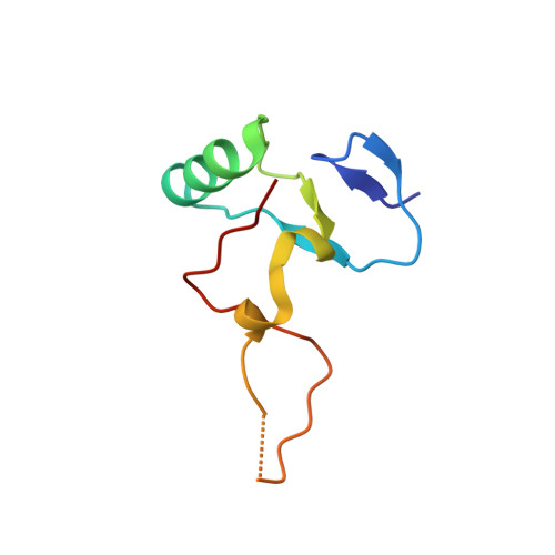
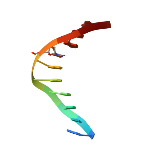
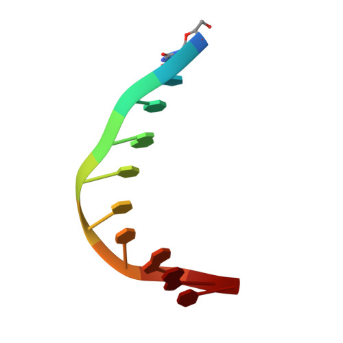
THAP proteins target specific DNA sites through bipartite recognition of adjacent major and minor grooves.

Sabogal, A.Lyubimov, A.Y.Corn, J.E.Berger, J.M.Rio, D.C.

(2010) Nat Struct Mol Biol 17: 117-123

  • DOI: https://doi.org/10.1038/nsmb.1742
  • Primary Citation of Related Structures:  
    3KDE

  • PubMed Abstract: 

    THAP-family C(2)CH zinc-coordinating DNA-binding proteins function in diverse eukaryotic cellular processes, such as transposition, transcriptional repression, stem-cell pluripotency, angiogenesis and neurological function. To determine the molecular basis for sequence-specific DNA recognition by THAP proteins, we solved the crystal structure of the Drosophila melanogaster P element transposase THAP domain (DmTHAP) in complex with a natural 10-base-pair site. In contrast to C(2)H(2) zinc fingers, DmTHAP docks a conserved beta-sheet into the major groove and a basic C-terminal loop into the adjacent minor groove. We confirmed specific protein-DNA interactions by mutagenesis and DNA-binding assays. Sequence analysis of natural and in vitro-selected binding sites suggests that several THAPs (DmTHAP and human THAP1 and THAP9) recognize a bipartite TXXGGGX(A/T) consensus motif; homology suggests THAP proteins bind DNA through a bipartite interaction. These findings reveal the conserved mechanisms by which THAP-family proteins engage specific chromosomal target elements.


  • Organizational Affiliation

    Department of Molecular and Cell Biology, University of California, Berkeley, Berkeley, California, USA.


Macromolecules

Find similar proteins by:  (by identity cutoff)  |  3D Structure
Entity ID: 3
MoleculeChains Sequence LengthOrganismDetailsImage
Transposable element P transposase77Drosophila melanogasterMutation(s): 0 
Gene Names: T
UniProt
Find proteins for Q7M3K2 (Drosophila melanogaster)
Explore Q7M3K2 
Go to UniProtKB:  Q7M3K2
Entity Groups  
Sequence Clusters30% Identity50% Identity70% Identity90% Identity95% Identity100% Identity
UniProt GroupQ7M3K2
Sequence Annotations
Expand
  • Reference Sequence

Find similar nucleic acids by:  Sequence   |   3D Structure  

Entity ID: 1
MoleculeChains LengthOrganismImage
5'-D(*GP*TP*TP*AP*AP*GP*(BRU)P*GP*GP*A)-3'10N/A
Sequence Annotations
Expand
  • Reference Sequence

Find similar nucleic acids by:  Sequence   |   3D Structure  

Entity ID: 2
MoleculeChains LengthOrganismImage
5'-D(*(BRU)P*CP*CP*AP*CP*TP*TP*AP*AP*C)-3'10N/A
Sequence Annotations
Expand
  • Reference Sequence
Small Molecules
Ligands 1 Unique
IDChains Name / Formula / InChI Key2D Diagram3D Interactions
ZN
Query on ZN

Download Ideal Coordinates CCD File 
D [auth C]ZINC ION
Zn
PTFCDOFLOPIGGS-UHFFFAOYSA-N
Experimental Data & Validation

Experimental Data

  • Method: X-RAY DIFFRACTION
  • Resolution: 1.74 Å
  • R-Value Free: 0.216 
  • R-Value Work: 0.177 
  • R-Value Observed: 0.179 
  • Space Group: P 1 21 1
Unit Cell:
Length ( Å )Angle ( ˚ )
a = 28.692α = 90
b = 69.325β = 92.52
c = 35.126γ = 90
Software Package:
Software NamePurpose
Blu-Icedata collection
PHENIXmodel building
REFMACrefinement
HKL-2000data reduction
HKL-2000data scaling
PHENIXphasing

Structure Validation

View Full Validation Report


Entry History 

Deposition Data

Revision History  (Full details and data files)

  • Version 1.0: 2009-12-08
    Type: Initial release
  • Version 1.1: 2011-07-13
    Changes: Version format compliance
  • Version 1.2: 2012-02-01
    Changes: Other
  • Version 1.3: 2012-03-21
    Changes: Database references
  • Version 1.4: 2021-10-13
    Changes: Database references, Derived calculations
  • Version 1.5: 2024-02-21
    Changes: Data collection