3ISO

Crystal structure of 26 kDa GST of Clonorchis sinensis in P3221 symmetry


Experimental Data Snapshot

  • Method: X-RAY DIFFRACTION
  • Resolution: 1.90 Å
  • R-Value Free: 0.226 
  • R-Value Work: 0.179 
  • R-Value Observed: 0.181 

wwPDB Validation   3D Report Full Report


Ligand Structure Quality Assessment 


This is version 1.3 of the entry. See complete history


Literature

A histidine substitution confers metal binding affinity to a Schistosoma japonicum Glutathione S-transferase.

Han, Y.H.Seo, H.A.Kim, G.H.Lee, C.K.Kang, Y.K.Ryu, K.H.Chung, Y.J.

(2010) Protein Expr Purif 73: 74-77

  • DOI: https://doi.org/10.1016/j.pep.2010.03.014
  • Primary Citation of Related Structures:  
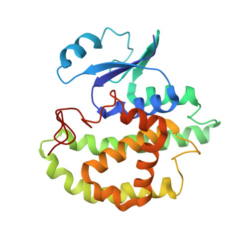
    3ISO

  • PubMed Abstract: 

    Glutathione S-transferases (GSTs) are multifunctional enzymes that are used as fusion tags on recombinant proteins in mammalian and Escherichia coli expression systems. We recently found that the Schistosoma japonicum GST (SjGST) displays weak Ni(2+) ion binding affinity. Glu26 and His79 were assumed to be its Ni(2+) binding sites based on the structure of the 26-kDa Clonorchis sinensis GST. To enhance SjGST Ni(2+) binding affinity, Glu26 was mutated to His. SjGST-E26H was expressed and purified at a high concentration of imidazole to a higher purity than wild type SjGST. In addition, human biotin protein ligase fused to SjGST-E26H was purified with a immobilized Ni affinity column.


  • Organizational Affiliation

    Department of Biochemistry, Chungbuk National University, Cheongju 361-763, Republic of Korea.


Macromolecules
Find similar proteins by:  (by identity cutoff)  |  3D Structure
Entity ID: 1
MoleculeChains Sequence LengthOrganismDetailsImage
Putative glutathione transferase
A, B
218Clonorchis sinensisMutation(s): 0 
UniProt
Find proteins for Q25595 (Clonorchis sinensis)
Explore Q25595 
Go to UniProtKB:  Q25595
Entity Groups  
Sequence Clusters30% Identity50% Identity70% Identity90% Identity95% Identity100% Identity
UniProt GroupQ25595
Sequence Annotations
Expand
  • Reference Sequence
Small Molecules
Ligands 3 Unique
IDChains Name / Formula / InChI Key2D Diagram3D Interactions
GSH
Query on GSH

Download Ideal Coordinates CCD File 
C [auth A],
J [auth B]
GLUTATHIONE
C10 H17 N3 O6 S
RWSXRVCMGQZWBV-WDSKDSINSA-N
SO4
Query on SO4

Download Ideal Coordinates CCD File 
E [auth A]
F [auth A]
G [auth A]
H [auth A]
I [auth A]
E [auth A],
F [auth A],
G [auth A],
H [auth A],
I [auth A],
L [auth B],
M [auth B],
N [auth B]
SULFATE ION
O4 S
QAOWNCQODCNURD-UHFFFAOYSA-L
ZN
Query on ZN

Download Ideal Coordinates CCD File 
D [auth A],
K [auth B]
ZINC ION
Zn
PTFCDOFLOPIGGS-UHFFFAOYSA-N
Experimental Data & Validation

Experimental Data

  • Method: X-RAY DIFFRACTION
  • Resolution: 1.90 Å
  • R-Value Free: 0.226 
  • R-Value Work: 0.179 
  • R-Value Observed: 0.181 
  • Space Group: P 32 2 1
Unit Cell:
Length ( Å )Angle ( ˚ )
a = 96.39α = 90
b = 96.39β = 90
c = 115.428γ = 120
Software Package:
Software NamePurpose
DENZOdata reduction
SCALEPACKdata scaling
MOLREPphasing
REFMACrefinement
PDB_EXTRACTdata extraction
HKL-2000data collection

Structure Validation

View Full Validation Report


Ligand Structure Quality Assessment 


Entry History 

Deposition Data

Revision History  (Full details and data files)

  • Version 1.0: 2010-09-08
    Type: Initial release
  • Version 1.1: 2011-07-13
    Changes: Version format compliance
  • Version 1.2: 2017-11-01
    Changes: Refinement description
  • Version 1.3: 2023-11-01
    Changes: Data collection, Database references, Derived calculations, Refinement description