3HD7

HELICAL EXTENSION OF THE NEURONAL SNARE COMPLEX INTO THE MEMBRANE, spacegroup C 1 2 1


Experimental Data Snapshot

  • Method: X-RAY DIFFRACTION
  • Resolution: 3.40 Å
  • R-Value Free: 0.271 
  • R-Value Work: 0.239 
  • R-Value Observed: 0.241 

wwPDB Validation   3D Report Full Report


Ligand Structure Quality Assessment 


This is version 1.3 of the entry. See complete history


Literature

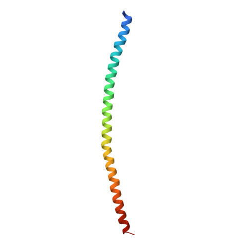
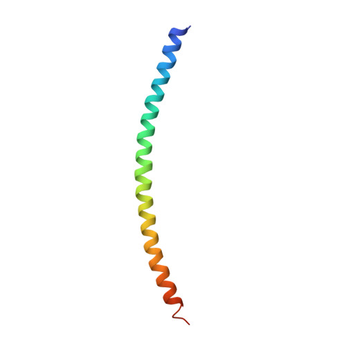
Helical extension of the neuronal SNARE complex into the membrane

Stein, A.Weber, G.Wahl, M.C.Jahn, R.

(2009) Nature 460: 525-528

  • DOI: https://doi.org/10.1038/nature08156
  • Primary Citation of Related Structures:  
    3HD7, 3IPD

  • PubMed Abstract: 

    Neurotransmission relies on synaptic vesicles fusing with the membrane of nerve cells to release their neurotransmitter content into the synaptic cleft, a process requiring the assembly of several members of the SNARE (soluble N-ethylmaleimide-sensitive factor attachment protein receptor) family. SNAREs represent an evolutionarily conserved protein family that mediates membrane fusion in the secretory and endocytic pathways of eukaryotic cells. On membrane contact, these proteins assemble in trans between the membranes as a bundle of four alpha-helices, with the energy released during assembly being thought to drive fusion. However, it is unclear how the energy is transferred to the membranes and whether assembly is conformationally linked to fusion. Here, we report the X-ray structure of the neuronal SNARE complex, consisting of rat syntaxin 1A, SNAP-25 and synaptobrevin 2, with the carboxy-terminal linkers and transmembrane regions at 3.4 A resolution. The structure shows that assembly proceeds beyond the already known core SNARE complex, resulting in a continuous helical bundle that is further stabilized by side-chain interactions in the linker region. Our results suggest that the final phase of SNARE assembly is directly coupled to membrane merger.


  • Organizational Affiliation

    Department of Neurobiology, Max Planck Institute for Biophysical Chemistry, 37077 Göttingen, Germany.


Macromolecules
Find similar proteins by:  (by identity cutoff)  |  3D Structure
Entity ID: 1
MoleculeChains Sequence LengthOrganismDetailsImage
Vesicle-associated membrane protein 2
A, E
91Rattus norvegicusMutation(s): 0 
Gene Names: Vamp2
Membrane Entity: Yes 
UniProt
Find proteins for P63045 (Rattus norvegicus)
Explore P63045 
Go to UniProtKB:  P63045
Entity Groups  
Sequence Clusters30% Identity50% Identity70% Identity90% Identity95% Identity100% Identity
UniProt GroupP63045
Sequence Annotations
Expand
  • Reference Sequence
Find similar proteins by:  (by identity cutoff)  |  3D Structure
Entity ID: 2
MoleculeChains Sequence LengthOrganismDetailsImage
Syntaxin-1A
B, F
109Rattus norvegicusMutation(s): 0 
Gene Names: Stx1a
Membrane Entity: Yes 
UniProt
Find proteins for P32851 (Rattus norvegicus)
Explore P32851 
Go to UniProtKB:  P32851
Entity Groups  
Sequence Clusters30% Identity50% Identity70% Identity90% Identity95% Identity100% Identity
UniProt GroupP32851
Sequence Annotations
Expand
  • Reference Sequence
Find similar proteins by:  (by identity cutoff)  |  3D Structure
Entity ID: 3
MoleculeChains Sequence LengthOrganismDetailsImage
Synaptosomal-associated protein 25
C, G
80Rattus norvegicusMutation(s): 0 
Gene Names: Snap25
Membrane Entity: Yes 
UniProt
Find proteins for P60881 (Rattus norvegicus)
Explore P60881 
Go to UniProtKB:  P60881
Entity Groups  
Sequence Clusters30% Identity50% Identity70% Identity90% Identity95% Identity100% Identity
UniProt GroupP60881
Sequence Annotations
Expand
  • Reference Sequence
Find similar proteins by:  (by identity cutoff)  |  3D Structure
Entity ID: 4
MoleculeChains Sequence LengthOrganismDetailsImage
Synaptosomal-associated protein 25
D, H
68Rattus norvegicusMutation(s): 0 
Gene Names: Snap25
Membrane Entity: Yes 
UniProt
Find proteins for P60881 (Rattus norvegicus)
Explore P60881 
Go to UniProtKB:  P60881
Entity Groups  
Sequence Clusters30% Identity50% Identity70% Identity90% Identity95% Identity100% Identity
UniProt GroupP60881
Sequence Annotations
Expand
  • Reference Sequence
Small Molecules
Ligands 2 Unique
IDChains Name / Formula / InChI Key2D Diagram3D Interactions
GGG
Query on GGG

Download Ideal Coordinates CCD File 
I [auth A],
J [auth A],
M [auth E]
glycylglycylglycine
C6 H11 N3 O4
XKUKSGPZAADMRA-UHFFFAOYSA-N
SO4
Query on SO4

Download Ideal Coordinates CCD File 
K [auth C]
L [auth D]
N [auth E]
O [auth E]
P [auth F]
K [auth C],
L [auth D],
N [auth E],
O [auth E],
P [auth F],
Q [auth F],
R [auth F],
S [auth H]
SULFATE ION
O4 S
QAOWNCQODCNURD-UHFFFAOYSA-L
Experimental Data & Validation

Experimental Data

  • Method: X-RAY DIFFRACTION
  • Resolution: 3.40 Å
  • R-Value Free: 0.271 
  • R-Value Work: 0.239 
  • R-Value Observed: 0.241 
  • Space Group: C 1 2 1
Unit Cell:
Length ( Å )Angle ( ˚ )
a = 265.412α = 90
b = 135.49β = 96.34
c = 58.689γ = 90
Software Package:
Software NamePurpose
SHELXSphasing
PHENIXrefinement
HKL-2000data reduction
HKL-2000data scaling

Structure Validation

View Full Validation Report


Ligand Structure Quality Assessment 


Entry History 

Deposition Data

Revision History  (Full details and data files)

  • Version 1.0: 2009-07-14
    Type: Initial release
  • Version 1.1: 2011-07-13
    Changes: Non-polymer description, Version format compliance
  • Version 1.2: 2023-11-01
    Changes: Data collection, Database references, Derived calculations, Refinement description
  • Version 1.3: 2024-03-20
    Changes: Source and taxonomy, Structure summary