3H9D

Crystal Structure of Trypanosoma brucei ATG8


Experimental Data Snapshot

  • Method: X-RAY DIFFRACTION
  • Resolution: 2.30 Å
  • R-Value Free: 0.310 
  • R-Value Work: 0.234 
  • R-Value Observed: 0.238 

wwPDB Validation   3D Report Full Report


This is version 1.2 of the entry. See complete history


Literature

Trypanosoma brucei ATG8: structural insights into autophagic-like mechanisms in protozoa.

Koopmann, R.Muhammad, K.Perbandt, M.Betzel, C.Duszenko, M.

(2009) Autophagy 5: 1085-1091

  • DOI: https://doi.org/10.4161/auto.5.8.9611
  • Primary Citation of Related Structures:  
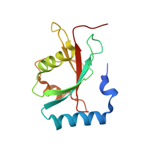
    3H9D

  • PubMed Abstract: 

    Bioinformatic searches of genome databases revealed that the number of autophagy-related genes (ATG) is considerably lower in trypanosomes than in higher eukaryotes and even in yeast. This raises the question of whether autophagy in this protozoan parasite is more primitive and represents a rudimentary paradigm due to its early branching off the evolutionary tree. We here present the crystal structure of TbATG8B. This molecule (MW 13.7 kDa) belongs to the ubiquitin-like proteins showing the typical ubiquitin fold and strong sequence homology to LC3, the human homologue. Due to its characteristic folding, it should readily bind to TbATG4.1 for being processed. This presumption was tested by molecular modeling approaches, docking TbATG8B to a homology model of TbATG4.1. Although exchanges of several amino acids are evident from sequence comparisons, the overall structure seems very much alike and the necessary catalytic triad (C-D-H) is well conserved in TbATG4.1. Thus membrane formation during appearance of the autophagic bodies seems very similar in trypanosomes and their higher eukaryotic counterpart.


  • Organizational Affiliation

    Interfaculty Institute for Biochemistry, University of Tübingen, Tübingen, Germany.


Macromolecules
Find similar proteins by:  (by identity cutoff)  |  3D Structure
Entity ID: 1
MoleculeChains Sequence LengthOrganismDetailsImage
Microtubule-associated protein 1A/1B, light chain 3, putative
A, B
119Trypanosoma bruceiMutation(s): 0 
UniProt
Find proteins for Q582K6 (Trypanosoma brucei brucei (strain 927/4 GUTat10.1))
Explore Q582K6 
Go to UniProtKB:  Q582K6
Entity Groups  
Sequence Clusters30% Identity50% Identity70% Identity90% Identity95% Identity100% Identity
UniProt GroupQ582K6
Sequence Annotations
Expand
  • Reference Sequence
Experimental Data & Validation

Experimental Data

  • Method: X-RAY DIFFRACTION
  • Resolution: 2.30 Å
  • R-Value Free: 0.310 
  • R-Value Work: 0.234 
  • R-Value Observed: 0.238 
  • Space Group: P 21 21 21
Unit Cell:
Length ( Å )Angle ( ˚ )
a = 48.1α = 90
b = 48.6β = 90
c = 127.92γ = 90
Software Package:
Software NamePurpose
REFMACrefinement

Structure Validation

View Full Validation Report


Entry History 

Deposition Data

Revision History  (Full details and data files)

  • Version 1.0: 2009-10-06
    Type: Initial release
  • Version 1.1: 2011-07-13
    Changes: Version format compliance
  • Version 1.2: 2024-02-21
    Changes: Data collection, Database references, Derived calculations