3GBE

Crystal structure of the isomaltulose synthase SmuA from Protaminobacter rubrum in complex with the inhibitor deoxynojirimycin


Experimental Data Snapshot

  • Method: X-RAY DIFFRACTION
  • Resolution: 1.70 Å
  • R-Value Free: 0.197 
  • R-Value Work: 0.171 
  • R-Value Observed: 0.171 

wwPDB Validation   3D Report Full Report


Ligand Structure Quality Assessment 


This is version 1.2 of the entry. See complete history


Literature

Structural determinants of product specificity of sucrose isomerases

Ravaud, S.Robert, X.Watzlawick, H.Haser, R.Mattes, R.Aghajari, N.

(2009) FEBS Lett 583: 1964-1968

  • DOI: https://doi.org/10.1016/j.febslet.2009.05.002
  • Primary Citation of Related Structures:  
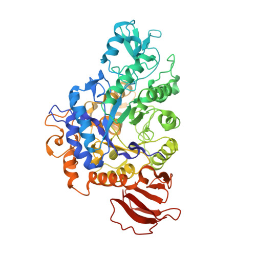
    3GBD, 3GBE

  • PubMed Abstract: 

    The healthy sweetener isomaltulose is industrially produced from the conversion of sucrose by the sucrose isomerase SmuA from Protaminobacter rubrum. Crystal structures of SmuA in native and deoxynojirimycin complexed forms completed with modeling studies unravel the characteristics of the isomaltulose synthases catalytic pocket and their substrate binding mode. Comparison with the trehalulose synthase MutB highlights the role of Arg(298) and Arg(306) active site residues and surface charges in controlling product specificity of sucrose isomerases (isomaltulose versus trehalulose). The results provide a rationale for the specific design of optimized enzymes.


  • Organizational Affiliation

    Institut de Biologie et Chimie des Protéines, UMR 5086-CNRS/Université de Lyon, IFR128 BioSciences Gerland-Lyon Sud, Lyon, France.


Macromolecules
Find similar proteins by:  (by identity cutoff)  |  3D Structure
Entity ID: 1
MoleculeChains Sequence LengthOrganismDetailsImage
Sucrose isomerase SmuA from Protaminobacter rubrum558Serratia plymuthicaMutation(s): 0 
Gene Names: smuA
EC: 5.4.99.11
UniProt
Find proteins for D0VX20 (Serratia plymuthica)
Explore D0VX20 
Go to UniProtKB:  D0VX20
Entity Groups  
Sequence Clusters30% Identity50% Identity70% Identity90% Identity95% Identity100% Identity
UniProt GroupD0VX20
Sequence Annotations
Expand
  • Reference Sequence
Small Molecules
Ligands 3 Unique
IDChains Name / Formula / InChI Key2D Diagram3D Interactions
FLC
Query on FLC

Download Ideal Coordinates CCD File 
J [auth A]CITRATE ANION
C6 H5 O7
KRKNYBCHXYNGOX-UHFFFAOYSA-K
NOJ
Query on NOJ

Download Ideal Coordinates CCD File 
K [auth A]1-DEOXYNOJIRIMYCIN
C6 H13 N O4
LXBIFEVIBLOUGU-JGWLITMVSA-N
EDO
Query on EDO

Download Ideal Coordinates CCD File 
B [auth A]
C [auth A]
D [auth A]
E [auth A]
F [auth A]
B [auth A],
C [auth A],
D [auth A],
E [auth A],
F [auth A],
G [auth A],
H [auth A],
I [auth A]
1,2-ETHANEDIOL
C2 H6 O2
LYCAIKOWRPUZTN-UHFFFAOYSA-N
Binding Affinity Annotations 
IDSourceBinding Affinity
NOJ Binding MOAD:  3GBE Ki: 1.00e+4 (nM) from 1 assay(s)
PDBBind:  3GBE Ki: 1.00e+4 (nM) from 1 assay(s)
Experimental Data & Validation

Experimental Data

  • Method: X-RAY DIFFRACTION
  • Resolution: 1.70 Å
  • R-Value Free: 0.197 
  • R-Value Work: 0.171 
  • R-Value Observed: 0.171 
  • Space Group: P 21 21 21
Unit Cell:
Length ( Å )Angle ( ˚ )
a = 60.9α = 90
b = 80.4β = 90
c = 135.83γ = 90
Software Package:
Software NamePurpose
ADSCdata collection
CNSrefinement
XDSdata reduction
XSCALEdata scaling
CNSphasing

Structure Validation

View Full Validation Report


Ligand Structure Quality Assessment 


Entry History 

Deposition Data

Revision History  (Full details and data files)

  • Version 1.0: 2009-05-26
    Type: Initial release
  • Version 1.1: 2011-07-13
    Changes: Non-polymer description, Version format compliance
  • Version 1.2: 2023-11-01
    Changes: Data collection, Database references, Derived calculations, Refinement description