3G5G

Crystal Structure of the Wild-Type Restriction-Modification Controller Protein C.Esp1396I


Experimental Data Snapshot

  • Method: X-RAY DIFFRACTION
  • Resolution: 2.80 Å
  • R-Value Free: 0.269 
  • R-Value Work: 0.237 
  • R-Value Observed: 0.239 

wwPDB Validation   3D Report Full Report


This is version 1.3 of the entry. See complete history


Literature

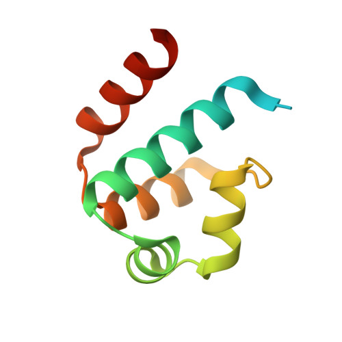
Structure of the restriction-modification controller protein C.Esp1396I.

Ball, N.Streeter, S.D.Kneale, G.G.McGeehan, J.E.

(2009) Acta Crystallogr D Biol Crystallogr 65: 900-905

  • DOI: https://doi.org/10.1107/S0907444909020514
  • Primary Citation of Related Structures:  
    3FYA, 3G5G

  • PubMed Abstract: 

    The controller protein of the Esp1396I restriction-modification (R-M) system binds differentially to three distinct operator sequences upstream of the methyltransferase (M) and endonuclease (R) genes to regulate the timing of gene expression. The crystal structure of a complex of the protein with two adjacent operator DNA sequences has been reported; however, the structure of the free protein has not yet been determined. Here, the crystal structure of the free protein is reported, with seven dimers in the asymmetric unit. Two of the 14 monomers show an alternative conformation to the major conformer in which the side chains of residues 43-46 in the loop region flanking the DNA-recognition helix are displaced by up to 10 A. It is proposed that the adoption of these two conformational states may play a role in DNA-sequence promiscuity. The two alternative conformations are also found in the R35A mutant structure, which is otherwise identical to the native protein. Comparison of the free and bound protein structures shows a 1.4 A displacement of the recognition helices when the dimer is bound to its DNA target.


  • Organizational Affiliation

    Biophysics Laboratories, Institute of Biomedical and Biomolecular Sciences, School of Biological Sciences, University of Portsmouth, Portsmouth, UK.


Macromolecules
Find similar proteins by:  (by identity cutoff)  |  3D Structure
Entity ID: 1
MoleculeChains Sequence LengthOrganismDetailsImage
Regulatory protein
A, B, C, D, E
A, B, C, D, E, F, G, H, I, J, K, L, M, N
99Enterobacter sp. RFL1396Mutation(s): 0 
Gene Names: esp1396IC
UniProt
Find proteins for Q8GGH0 (Enterobacter sp. RFL1396)
Explore Q8GGH0 
Go to UniProtKB:  Q8GGH0
Entity Groups  
Sequence Clusters30% Identity50% Identity70% Identity90% Identity95% Identity100% Identity
UniProt GroupQ8GGH0
Sequence Annotations
Expand
  • Reference Sequence
Experimental Data & Validation

Experimental Data

  • Method: X-RAY DIFFRACTION
  • Resolution: 2.80 Å
  • R-Value Free: 0.269 
  • R-Value Work: 0.237 
  • R-Value Observed: 0.239 
  • Space Group: P 65
Unit Cell:
Length ( Å )Angle ( ˚ )
a = 128.717α = 90
b = 128.717β = 90
c = 137.508γ = 120
Software Package:
Software NamePurpose
XSCALEdata scaling
PHASERphasing
REFMACrefinement
PDB_EXTRACTdata extraction
MxCuBEdata collection
XDSdata reduction

Structure Validation

View Full Validation Report


Entry History 

Deposition Data

Revision History  (Full details and data files)

  • Version 1.0: 2009-08-25
    Type: Initial release
  • Version 1.1: 2011-07-13
    Changes: Advisory, Source and taxonomy, Version format compliance
  • Version 1.2: 2012-03-28
    Changes: Database references
  • Version 1.3: 2024-02-21
    Changes: Data collection, Database references, Refinement description