3FOU

Low pH structure of the Rieske protein from Thermus thermophilus at 2.1 A


Experimental Data Snapshot

  • Method: X-RAY DIFFRACTION
  • Resolution: 2.10 Å
  • R-Value Free: 0.246 
  • R-Value Work: 0.198 
  • R-Value Observed: 0.201 

wwPDB Validation   3D Report Full Report


This is version 1.2 of the entry. See complete history


Literature

Effects of pH on the Rieske Protein from Thermus thermophilus: A Spectroscopic and Structural Analysis

Konkle, M.E.Muellner, S.K.Schwander, A.L.Dicus, M.M.Pokhrel, R.Britt, R.D.Taylor, A.B.Hunsicker-Wang, L.M.

(2009) Biochemistry 48: 9848-9857

  • DOI: https://doi.org/10.1021/bi901126u
  • Primary Citation of Related Structures:  
    3FOU

  • PubMed Abstract: 

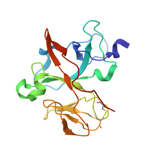
    The Rieske protein from Thermus thermophilus (TtRp) and a truncated version of the protein (truncTtRp), produced to achieve a low-pH crystallization condition, have been characterized using UV-visible and circular dichroism spectroscopies. TtRp and truncTtRp undergo a change in the UV-visible spectra with increasing pH. The LMCT band at 458 nm shifts to 436 nm and increases in intensity. The increase at 436 nm versus pH can be fit using the sum of two Henderson-Hasselbalch equations, yielding two pK(a) values for the oxidized protein. For TtRp, pK(ox1) = 7.48 +/- 0.12 and pK(ox2) = 10.07 +/- 0.17. For truncTtRp, pK(ox1) = 7.87 +/- 0.17 and pK(ox2) = 9.84 +/- 0.42. The shift to shorter wavelength and the increase in intensity for the LMCT band with increasing pH are consistent with deprotonation of the histidine ligands. A pH titration of truncTtRp monitored by circular dichroism also showed pH-dependent changes at 315 and 340 nm. At 340 nm, the fit gives pK(ox1) = 7.14 +/- 0.26 and pK(ox2) = 9.32 +/- 0.36. The change at 315 nm is best fit for a single deprotonation event, giving pK(ox1) = 7.82 +/- 0.10. The lower wavelength region of the CD spectra was unaffected by pH, indicating that the overall fold of the protein remains unchanged, which is consistent with crystallographic results of truncTtRp. The structure of truncTtRp crystallized at pH 6.2 is very similar to TtRp at pH 8.5 and contains only subtle changes localized at the [2Fe-2S] cluster. These titration and structural results further elucidate the histidine ligand characteristics and are consistent with important roles for these amino acids.


  • Organizational Affiliation

    Department of Chemistry, Trinity University, One Trinity Place, San Antonio, Texas 78212, USA.


Macromolecules
Find similar proteins by:  (by identity cutoff)  |  3D Structure
Entity ID: 1
MoleculeChains Sequence LengthOrganismDetailsImage
Quinol-cytochrome c reductase, Rieske iron-sulfur subunit
A, B
156Thermus thermophilus HB8Mutation(s): 0 
Gene Names: TTHA1931
UniProt
Find proteins for Q5SGZ9 (Thermus thermophilus (strain ATCC 27634 / DSM 579 / HB8))
Explore Q5SGZ9 
Go to UniProtKB:  Q5SGZ9
Entity Groups  
Sequence Clusters30% Identity50% Identity70% Identity90% Identity95% Identity100% Identity
UniProt GroupQ5SGZ9
Sequence Annotations
Expand
  • Reference Sequence
Small Molecules
Ligands 4 Unique
IDChains Name / Formula / InChI Key2D Diagram3D Interactions
FES
Query on FES

Download Ideal Coordinates CCD File 
C [auth A],
L [auth B]
FE2/S2 (INORGANIC) CLUSTER
Fe2 S2
NIXDOXVAJZFRNF-UHFFFAOYSA-N
PR
Query on PR

Download Ideal Coordinates CCD File 
D [auth A]
E [auth A]
F [auth A]
G [auth A]
H [auth A]
D [auth A],
E [auth A],
F [auth A],
G [auth A],
H [auth A],
M [auth B]
PRASEODYMIUM ION
Pr
WCWKKSOQLQEJTE-UHFFFAOYSA-N
ACT
Query on ACT

Download Ideal Coordinates CCD File 
J [auth A],
K [auth A]
ACETATE ION
C2 H3 O2
QTBSBXVTEAMEQO-UHFFFAOYSA-M
CA
Query on CA

Download Ideal Coordinates CCD File 
I [auth A],
N [auth B]
CALCIUM ION
Ca
BHPQYMZQTOCNFJ-UHFFFAOYSA-N
Experimental Data & Validation

Experimental Data

  • Method: X-RAY DIFFRACTION
  • Resolution: 2.10 Å
  • R-Value Free: 0.246 
  • R-Value Work: 0.198 
  • R-Value Observed: 0.201 
  • Space Group: P 21 21 2
Unit Cell:
Length ( Å )Angle ( ˚ )
a = 113.152α = 90
b = 58.512β = 90
c = 58.798γ = 90
Software Package:
Software NamePurpose
HKL-2000data collection
MOLREPphasing
REFMACrefinement
HKL-2000data reduction
HKL-2000data scaling

Structure Validation

View Full Validation Report


Entry History 

Deposition Data

Revision History  (Full details and data files)

  • Version 1.0: 2009-10-20
    Type: Initial release
  • Version 1.1: 2011-07-13
    Changes: Source and taxonomy, Version format compliance
  • Version 1.2: 2023-09-06
    Changes: Data collection, Database references, Derived calculations, Refinement description