3AQJ

Crystal Structure of a C-terminal domain of the bacteriophage P2 tail spike protein, gpV


Experimental Data Snapshot

  • Method: X-RAY DIFFRACTION
  • Resolution: 1.27 Å
  • R-Value Free: 0.190 
  • R-Value Work: 0.160 
  • R-Value Observed: 0.162 

wwPDB Validation   3D Report Full Report


This is version 1.2 of the entry. See complete history


Literature

The host-binding domain of the P2 phage tail spike reveals a trimeric iron-binding structure

Yamashita, E.Nakagawa, A.Takahashi, J.Tsunoda, K.Yamada, S.Takeda, S.

(2011) Acta Crystallogr Sect F Struct Biol Cryst Commun 67: 837-841

  • DOI: https://doi.org/10.1107/S1744309111005999
  • Primary Citation of Related Structures:  
    3AQJ

  • PubMed Abstract: 

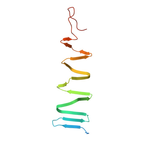
    The adsorption and infection of bacteriophage P2 is mediated by tail fibres and tail spikes. The tail spikes on the tail baseplate are used to irreversibly adsorb to the host cells. Recently, a P2 phage tail-spike protein, gpV, was purified and it was shown that a C-terminal domain, Ser87-Leu211, is sufficient for the binding of gpV to host Escherichia coli membranes [Kageyama et al. (2009), Biochemistry, 48, 10129-10135]. In this paper, the crystal structure of the C-terminal domain of P2 gpV is reported. The structure is a triangular pyramid and looks like a spearhead composed of an intertwined β-sheet, a triple β-helix and a metal-binding region containing iron, calcium and chloride ions.


  • Organizational Affiliation

    Institute for Protein Research, Osaka University, Suita, Osaka, Japan.


Macromolecules
Find similar proteins by:  (by identity cutoff)  |  3D Structure
Entity ID: 1
MoleculeChains Sequence LengthOrganismDetailsImage
Baseplate assembly protein V134Peduovirus P2Mutation(s): 0 
Gene Names: V
UniProt
Find proteins for P31340 (Escherichia phage P2)
Explore P31340 
Go to UniProtKB:  P31340
Entity Groups  
Sequence Clusters30% Identity50% Identity70% Identity90% Identity95% Identity100% Identity
UniProt GroupP31340
Sequence Annotations
Expand
  • Reference Sequence
Experimental Data & Validation

Experimental Data

  • Method: X-RAY DIFFRACTION
  • Resolution: 1.27 Å
  • R-Value Free: 0.190 
  • R-Value Work: 0.160 
  • R-Value Observed: 0.162 
  • Space Group: P 1 21 1
Unit Cell:
Length ( Å )Angle ( ˚ )
a = 66.912α = 90
b = 64.463β = 91.32
c = 67.186γ = 90
Software Package:
Software NamePurpose
SHARPphasing
REFMACrefinement
HKL-2000data reduction
HKL-2000data scaling

Structure Validation

View Full Validation Report


Entry History 

Deposition Data

Revision History  (Full details and data files)

  • Version 1.0: 2011-08-10
    Type: Initial release
  • Version 1.1: 2012-04-25
    Changes: Database references
  • Version 1.2: 2024-03-13
    Changes: Data collection, Database references, Derived calculations