Structural and functional analysis of three beta-glucosidases from bacterium Clostridium cellulovorans, fungus Trichoderma reesei and termite Neotermes koshunensis
Jeng, W.-Y., Wang, N.-C., Lin, M.-H., Lin, C.-T., Liaw, Y.-C., Chang, W.-J., Liu, C.-I., Liang, P.-H., Wang, A.H.-J.(2011) J Struct Biol 173: 46-56
- PubMed: 20682343
- DOI: https://doi.org/10.1016/j.jsb.2010.07.008
- Primary Citation of Related Structures:
3AHX, 3AHY, 3AHZ, 3AI0 - PubMed Abstract:
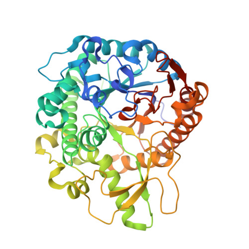
β-glucosidases (EC 3.2.1.21) cleave β-glucosidic linkages in disaccharide or glucose-substituted molecules and play important roles in fundamental biological processes. β-Glucosidases have been widely used in agricultural, biotechnological, industrial and medical applications. In this study, a high yield expression (70-250 mg/l) in Escherichia coli of the three functional β-glucosidase genes was obtained from the bacterium Clostridium cellulovorans (CcBglA), the fungus Trichoderma reesei (TrBgl2), and the termite Neotermes koshunensis (NkBgl) with the crystal structures of CcBglA, TrBgl2 and NkBgl, determined at 1.9Å, 1.63Å and 1.34Å resolution, respectively. The overall structures of these enzymes are similar to those belonging to the β-retaining glycosyl hydrolase family 1, which have a classical (α/β)(8)-TIM barrel fold. Each contains a slot-like active site cleft and a more variable outer opening, related to its function in processing different lengths of β-1,4-linked glucose derivatives. The two essential glutamate residues for hydrolysis are spatially conserved in the active site. In both TrBgl2 and NkBgl structures, a Tris molecule was found to bind at the active site, explaining the slight inhibition of hydrolase activity observed in Tris buffer. Manganese ions at 10mM exerted an approximate 2-fold enzyme activity enhancement of all three β-glucosidases, with CcBglA catalyzing the most efficiently in hydrolysis reaction and tolerating Tris as well as some metal inhibition. In summary, our results for the structural and functional properties of these three β-glucosidases from various biological sources open important avenues of exploration for further practical applications.
Organizational Affiliation:
Institute of Biological Chemistry, Academia Sinica, Taipei 115, Taiwan.