


Crystal structure of human Tob1 protein
Saito, K., Kishishita, S., Nishino, A., Murayama, K., Terada, T., Shirouzu, M., Kigawa, T., Yokoyama, S.To be published.
Experimental Data Snapshot
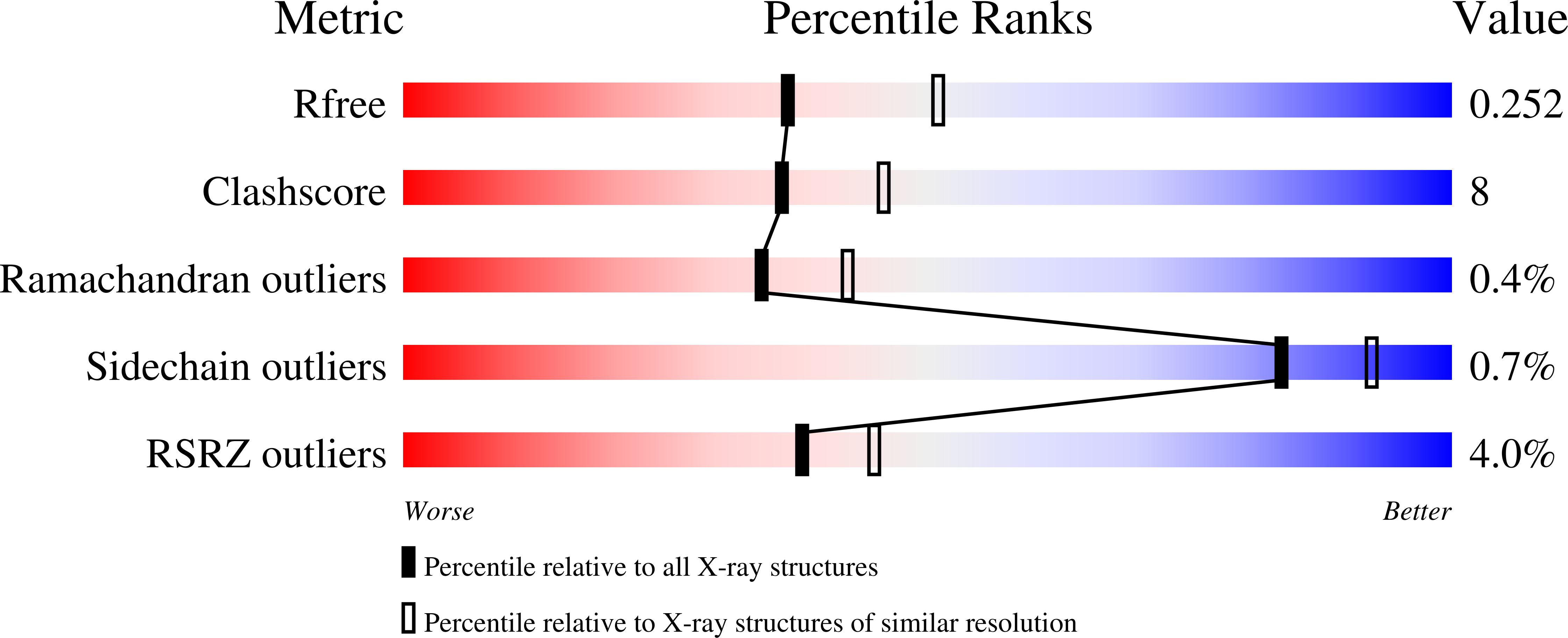
wwPDB Validation 3D Report Full Report
Entity ID: 1 | |||||
|---|---|---|---|---|---|
| Molecule | Chains | Sequence Length | Organism | Details | Image |
| Protein Tob1 | 130 | Homo sapiens | Mutation(s): 0 Gene Names: TOB1, TOB, TROB1 | 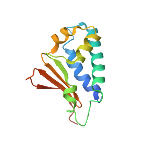 | |
UniProt & NIH Common Fund Data Resources | |||||
Find proteins for P50616 (Homo sapiens) Explore P50616 Go to UniProtKB: P50616 | |||||
PHAROS: P50616 GTEx: ENSG00000141232 | |||||
Entity Groups | |||||
| Sequence Clusters | 30% Identity50% Identity70% Identity90% Identity95% Identity100% Identity | ||||
| UniProt Group | P50616 | ||||
Sequence AnnotationsExpand | |||||
| |||||
| Length ( Å ) | Angle ( ˚ ) |
|---|---|
| a = 36.064 | α = 90 |
| b = 121.162 | β = 90 |
| c = 152.553 | γ = 90 |
| Software Name | Purpose |
|---|---|
| CNS | refinement |
| HKL-2000 | data reduction |
| HKL-2000 | data scaling |
| SOLVE | phasing |
RCSB PDB (citation) is hosted by
RCSB PDB is a member of the

