2XYP

Caspase-3:CAS26049945


Experimental Data Snapshot

  • Method: X-RAY DIFFRACTION
  • Resolution: 1.86 Å
  • R-Value Free: 0.204 
  • R-Value Work: 0.167 
  • R-Value Observed: 0.167 

wwPDB Validation   3D Report Full Report


Ligand Structure Quality Assessment 


This is version 1.1 of the entry. See complete history


Literature

In Silico Identification and Crystal Structure Validation of Caspase-3 Inhibitors without a P1 Aspartic Acid Moiety.

Ganesan, R.Jelakovic, S.Mittl, P.R.Caflisch, A.Grutter, M.G.

(2011) Acta Crystallogr Sect F Struct Biol Cryst Commun 67: 842

  • DOI: https://doi.org/10.1107/S1744309111018604
  • Primary Citation of Related Structures:  
    2XYG, 2XYH, 2XYP

  • PubMed Abstract: 

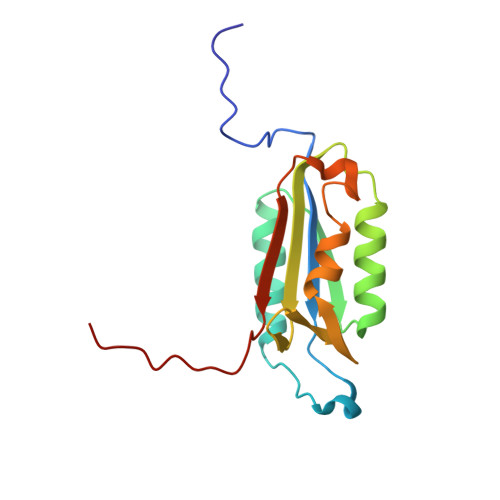
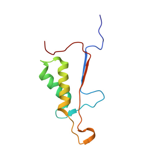
    Using a fragment-based docking procedure, several small-molecule inhibitors of caspase-3 were identified and tested and the crystal structures of three inhibitor complexes were determined. The crystal structures revealed that one inhibitor (NSC 18508) occupies only the S1 subsite, while two other inhibitors (NSC 89167 and NSC 251810) bind only to the prime part of the substrate-binding site. One of the major conformational changes observed in all three caspase-3-inhibitor complexes is a rotation of the Tyr204 side chain, which blocks the S2 subsite. In addition, the structural variability of the residues shaping the S1-S4 as well as the S1' subsites supports an induced-fit mechanism for the binding of the inhibitors in the active site. The high-resolution crystal structures reported here provide novel insights into the architecture of the substrate-binding site, which might be useful for the design of more potent caspase inhibitors.


  • Organizational Affiliation

    Department of Biochemistry, University of Zürich, Zürich, Switzerland.


Macromolecules
Find similar proteins by:  (by identity cutoff)  |  3D Structure
Entity ID: 1
MoleculeChains Sequence LengthOrganismDetailsImage
CASPASE-3 SUBUNIT P17146Homo sapiensMutation(s): 0 
EC: 3.4.22.56
UniProt & NIH Common Fund Data Resources
Find proteins for P42574 (Homo sapiens)
Explore P42574 
Go to UniProtKB:  P42574
PHAROS:  P42574
GTEx:  ENSG00000164305 
Entity Groups  
Sequence Clusters30% Identity50% Identity70% Identity90% Identity95% Identity100% Identity
UniProt GroupP42574
Sequence Annotations
Expand
  • Reference Sequence
Find similar proteins by:  (by identity cutoff)  |  3D Structure
Entity ID: 2
MoleculeChains Sequence LengthOrganismDetailsImage
CASPASE-3 SUBUNIT P1293Homo sapiensMutation(s): 0 
EC: 3.4.22.56
UniProt & NIH Common Fund Data Resources
Find proteins for P42574 (Homo sapiens)
Explore P42574 
Go to UniProtKB:  P42574
PHAROS:  P42574
GTEx:  ENSG00000164305 
Entity Groups  
Sequence Clusters30% Identity50% Identity70% Identity90% Identity95% Identity100% Identity
UniProt GroupP42574
Sequence Annotations
Expand
  • Reference Sequence
Small Molecules
Ligands 1 Unique
IDChains Name / Formula / InChI Key2D Diagram3D Interactions
XVE
Query on XVE

Download Ideal Coordinates CCD File 
C [auth A]PHENYLMETHYL N-[(2S)-4-CHLORO-3-OXO-1-PHENYL-BUTAN-2-YL]CARBAMATE
C18 H18 Cl N O3
OYHLRJGDELITAF-INIZCTEOSA-N
Experimental Data & Validation

Experimental Data

  • Method: X-RAY DIFFRACTION
  • Resolution: 1.86 Å
  • R-Value Free: 0.204 
  • R-Value Work: 0.167 
  • R-Value Observed: 0.167 
  • Space Group: I 2 2 2
Unit Cell:
Length ( Å )Angle ( ˚ )
a = 69.569α = 90
b = 83.782β = 90
c = 95.805γ = 90
Software Package:
Software NamePurpose
CNSrefinement

Structure Validation

View Full Validation Report


Ligand Structure Quality Assessment 


Entry History 

Deposition Data

Revision History  (Full details and data files)

  • Version 1.0: 2011-08-17
    Type: Initial release
  • Version 1.1: 2017-06-28
    Changes: Data collection