2UY7

Crystal structure of the P pilus rod subunit PapA


Experimental Data Snapshot

  • Method: X-RAY DIFFRACTION
  • Resolution: 2.60 Å
  • R-Value Free: 0.266 
  • R-Value Work: 0.230 
  • R-Value Observed: 0.230 

wwPDB Validation   3D Report Full Report


This is version 1.3 of the entry. See complete history


Literature

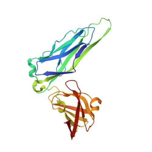
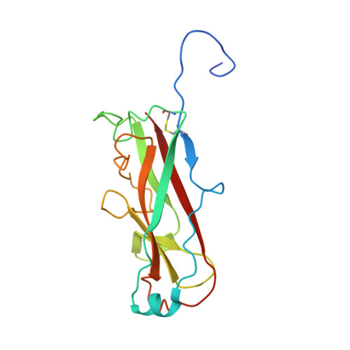
Crystal Structure of the P Pilus Rod Subunit Papa.

Verger, D.Bullitt, E.Hultgren, S.J.Waksman, G.

(2007) PLoS Pathog 3: E73

  • DOI: https://doi.org/10.1371/journal.ppat.0030073
  • Primary Citation of Related Structures:  
    2UY6, 2UY7

  • PubMed Abstract: 

    P pili are important adhesive fibres involved in kidney infection by uropathogenic Escherichia coli strains. P pili are assembled by the conserved chaperone-usher pathway, which involves the PapD chaperone and the PapC usher. During pilus assembly, subunits are incorporated into the growing fiber via the donor-strand exchange (DSE) mechanism, whereby the chaperone's G1 beta-strand that complements the incomplete immunoglobulin-fold of each subunit is displaced by the N-terminal extension (Nte) of an incoming subunit. P pili comprise a helical rod, a tip fibrillum, and an adhesin at the distal end. PapA is the rod subunit and is assembled into a superhelical right-handed structure. Here, we have solved the structure of a ternary complex of PapD bound to PapA through donor-strand complementation, itself bound to another PapA subunit through DSE. This structure provides insight into the structural basis of the DSE reaction involving this important pilus subunit. Using gel filtration chromatography and electron microscopy on a number of PapA Nte mutants, we establish that PapA differs in its mode of assembly compared with other Pap subunits, involving a much larger Nte that encompasses not only the DSE region of the Nte but also the region N-terminal to it.


  • Organizational Affiliation

    Institute of Structural Molecular Biology, University College London and Birkbeck College, London, United Kingdom.


Macromolecules
Find similar proteins by:  (by identity cutoff)  |  3D Structure
Entity ID: 1
MoleculeChains Sequence LengthOrganismDetailsImage
PERIPLASMID CHAPERONE PAPD PROTEIN
A, C, E, G
218Escherichia coli UTI89Mutation(s): 0 
UniProt
Find proteins for Q1R2W9 (Escherichia coli (strain UTI89 / UPEC))
Explore Q1R2W9 
Go to UniProtKB:  Q1R2W9
Entity Groups  
Sequence Clusters30% Identity50% Identity70% Identity90% Identity95% Identity100% Identity
UniProt GroupQ1R2W9
Sequence Annotations
Expand
  • Reference Sequence
Find similar proteins by:  (by identity cutoff)  |  3D Structure
Entity ID: 2
MoleculeChains Sequence LengthOrganismDetailsImage
PAP FIMBRIAL MAJOR PILIN PROTEIN
B, D, F, H
163Escherichia coliMutation(s): 1 
UniProt
Find proteins for P04127 (Escherichia coli)
Explore P04127 
Go to UniProtKB:  P04127
Entity Groups  
Sequence Clusters30% Identity50% Identity70% Identity90% Identity95% Identity100% Identity
UniProt GroupP04127
Sequence Annotations
Expand
  • Reference Sequence
Small Molecules
Ligands 1 Unique
IDChains Name / Formula / InChI Key2D Diagram3D Interactions
SO4
Query on SO4

Download Ideal Coordinates CCD File 
I [auth A]
J [auth A]
K [auth A]
L [auth A]
M [auth A]
I [auth A],
J [auth A],
K [auth A],
L [auth A],
M [auth A],
N [auth C],
O [auth C],
P [auth C],
Q [auth E],
R [auth G],
S [auth G],
T [auth G],
U [auth G]
SULFATE ION
O4 S
QAOWNCQODCNURD-UHFFFAOYSA-L
Experimental Data & Validation

Experimental Data

  • Method: X-RAY DIFFRACTION
  • Resolution: 2.60 Å
  • R-Value Free: 0.266 
  • R-Value Work: 0.230 
  • R-Value Observed: 0.230 
  • Space Group: P 32 2 1
Unit Cell:
Length ( Å )Angle ( ˚ )
a = 166.96α = 90
b = 166.96β = 90
c = 178.01γ = 120
Software Package:
Software NamePurpose
CNSrefinement
MOSFLMdata reduction
SCALAdata scaling
AMoREphasing

Structure Validation

View Full Validation Report


Entry History 

Deposition Data

Revision History  (Full details and data files)

  • Version 1.0: 2007-05-29
    Type: Initial release
  • Version 1.1: 2011-05-08
    Changes: Version format compliance
  • Version 1.2: 2011-07-13
    Changes: Version format compliance
  • Version 1.3: 2023-12-13
    Changes: Data collection, Database references, Other, Refinement description