2QFJ

Crystal Structure of First Two RRM Domains of FIR Bound to ssDNA from a Portion of FUSE


Experimental Data Snapshot

  • Method: X-RAY DIFFRACTION
  • Resolution: 2.10 Å
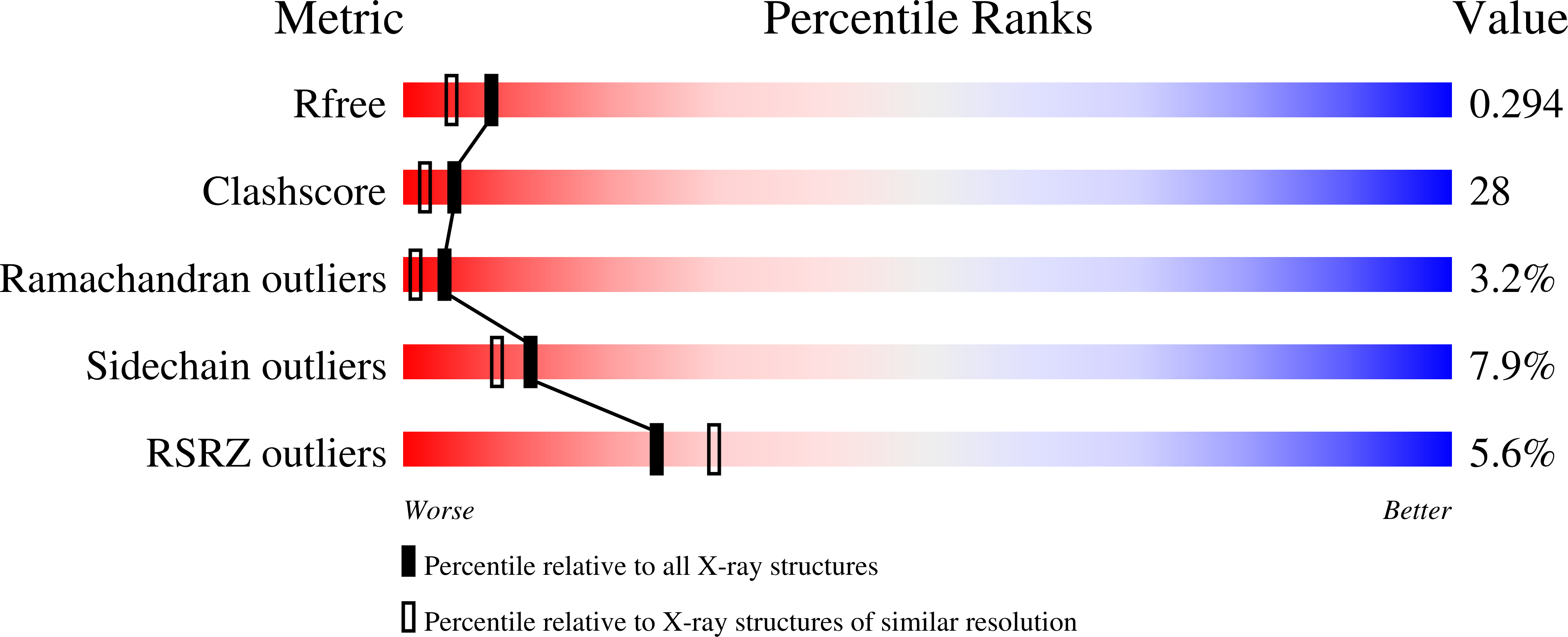
  • R-Value Free: 0.295 
  • R-Value Work: 0.258 
  • R-Value Observed: 0.258 

wwPDB Validation   3D Report Full Report


This is version 1.4 of the entry. See complete history


Literature

Dimerization of FIR upon FUSE DNA binding suggests a mechanism of c-myc inhibition

Crichlow, G.V.Zhou, H.Hsiao, H.-H.Frederick, K.B.Debrosse, M.Yang, Y.Folta-Stogniew, E.J.Chung, H.-J.Fan, C.De La Cruz, E.M.Levens, D.Lolis, E.Braddock, D.

(2007) EMBO J 27: 277-289

  • DOI: https://doi.org/10.1038/sj.emboj.7601936
  • Primary Citation of Related Structures:  
    2QFJ

  • PubMed Abstract: 

    c-myc is essential for cell homeostasis and growth but lethal if improperly regulated. Transcription of this oncogene is governed by the counterbalancing forces of two proteins on TFIIH--the FUSE binding protein (FBP) and the FBP-interacting repressor (FIR). FBP and FIR recognize single-stranded DNA upstream of the P1 promoter, known as FUSE, and influence transcription by oppositely regulating TFIIH at the promoter site. Size exclusion chromatography coupled with light scattering reveals that an FIR dimer binds one molecule of single-stranded DNA. The crystal structure confirms that FIR binds FUSE as a dimer, and only the N-terminal RRM domain participates in nucleic acid recognition. Site-directed mutations of conserved residues in the first RRM domain reduce FIR's affinity for FUSE, while analogous mutations in the second RRM domain either destabilize the protein or have no effect on DNA binding. Oppositely oriented DNA on parallel binding sites of the FIR dimer results in spooling of a single strand of bound DNA, and suggests a mechanism for c-myc transcriptional control.


  • Organizational Affiliation

    Department of Pharmacology, Yale University School of Medicine, New Haven, CT 06510, USA.


Macromolecules

Find similar proteins by:  (by identity cutoff)  |  3D Structure
Entity ID: 2
MoleculeChains Sequence LengthOrganismDetailsImage
FBP-interacting repressorB [auth A],
C [auth B]
216Homo sapiensMutation(s): 3 
Gene Names: FIRSIAHBP1
UniProt & NIH Common Fund Data Resources
Find proteins for Q9UHX1 (Homo sapiens)
Explore Q9UHX1 
Go to UniProtKB:  Q9UHX1
PHAROS:  Q9UHX1
GTEx:  ENSG00000179950 
Entity Groups  
Sequence Clusters30% Identity50% Identity70% Identity90% Identity95% Identity100% Identity
UniProt GroupQ9UHX1
Sequence Annotations
Expand
  • Reference Sequence

Find similar nucleic acids by:  Sequence   |   3D Structure  

Entity ID: 1
MoleculeChains LengthOrganismImage
DNA (5'-D(*DTP*DCP*DGP*DGP*DGP*DAP*DTP*DTP*DTP*DTP*DTP*DTP*DAP*DTP*DTP*DTP*DTP*DGP*DTP*DGP*DTP*DTP*DAP*DTP*DT)-3')A [auth C]25N/A
Sequence Annotations
Expand
  • Reference Sequence
Experimental Data & Validation

Experimental Data

  • Method: X-RAY DIFFRACTION
  • Resolution: 2.10 Å
  • R-Value Free: 0.295 
  • R-Value Work: 0.258 
  • R-Value Observed: 0.258 
  • Space Group: P 31
Unit Cell:
Length ( Å )Angle ( ˚ )
a = 63.135α = 90
b = 63.135β = 90
c = 82.589γ = 120
Software Package:
Software NamePurpose
DENZOdata reduction
SCALEPACKdata scaling
AMoREphasing
CNSrefinement
PDB_EXTRACTdata extraction
HKL-2000data reduction
HKL-2000data scaling
SOLVEphasing

Structure Validation

View Full Validation Report


Entry History 

Deposition Data

Revision History  (Full details and data files)

  • Version 1.0: 2008-03-04
    Type: Initial release
  • Version 1.1: 2011-07-13
    Changes: Version format compliance
  • Version 1.2: 2017-10-18
    Changes: Refinement description
  • Version 1.3: 2021-10-20
    Changes: Database references
  • Version 1.4: 2024-02-21
    Changes: Data collection