2PRJ

Binding of N-acetyl-beta-D-glucopyranosylamine to Glycogen Phosphorylase B


Experimental Data Snapshot

  • Method: X-RAY DIFFRACTION
  • Resolution: 2.30 Å
  • R-Value Free: 0.237 
  • R-Value Work: 0.181 
  • R-Value Observed: 0.181 

wwPDB Validation   3D Report Full Report


This is version 1.5 of the entry. See complete history


Literature

N-acetyl-beta-D-glucopyranosylamine: a potent T-state inhibitor of glycogen phosphorylase. A comparison with alpha-D-glucose.

Oikonomakos, N.G.Kontou, M.Zographos, S.E.Watson, K.A.Johnson, L.N.Bichard, C.J.Fleet, G.W.Acharya, K.R.

(1995) Protein Sci 4: 2469-2477

  • DOI: https://doi.org/10.1002/pro.5560041203
  • Primary Citation of Related Structures:  
    2PRJ

  • PubMed Abstract: 

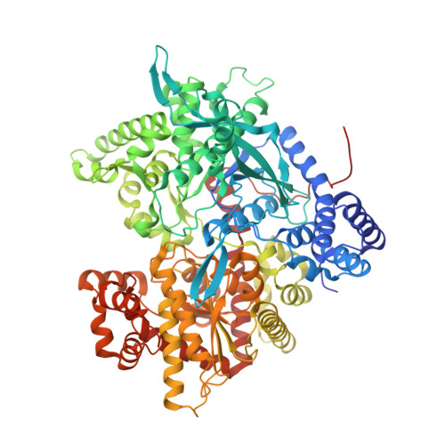
    Structure-based drug design has led to the discovery of a number of glucose analogue inhibitors of glycogen phosphorylase that have an increased affinity compared to alpha-D-glucose (Ki = 1.7 mM). The best inhibitor in the class of N-acyl derivatives of beta-D-glucopyranosylamine, N-acetyl-beta-D-glucopyranosylamine (1-GlcNAc), has been characterized by kinetic, ultracentrifugation, and crystallographic studies. 1-GlcNAc acts as a competitive inhibitor for both the b (Ki = 32 microM) and the a (Ki = 35 microM) forms of the enzyme with respect to glucose 1-phosphate and in synergism with caffeine, mimicking the binding of glucose. Sedimentation velocity experiments demonstrated that 1-GlcNAc was able to induce dissociation of tetrameric phosphorylase a and stabilization of the dimeric T-state conformation. Co-crystals of the phosphorylase b-1-GlcNAc-IMP complex were grown in space group P4(3)2(1)2, with native-like unit cell dimensions, and the complex structure has been refined to give a crystallographic R factor of 18.1%, for data between 8 and 2.3 A resolution. 1-GlcNAc binds tightly at the catalytic site of T-state phosphorylase b at approximately the same position as that of alpha-D-glucose. The ligand can be accommodated in the catalytic site with very little change in the protein structure and stabilizes the T-state conformation of the 280s loop by making several favorable contacts to Asn 284 of this loop. Structural comparisons show that the T-state phosphorylase b-1-GlcNAc-IMP complex structure is overall similar to the T-state phosphorylase b-alpha-D-glucose complex structure. The structure of the 1-GlcNAc complex provides a rational for the biochemical properties of the inhibitor.


  • Organizational Affiliation

    Institute of Biological Research and Biotechnology, National Hellenic Research Foundation, Athens, Greece. nikos@apollon.servicenet.ariadne.t.gr


Macromolecules
Find similar proteins by:  (by identity cutoff)  |  3D Structure
Entity ID: 1
MoleculeChains Sequence LengthOrganismDetailsImage
GLYCOGEN PHOSPHORYLASE842Oryctolagus cuniculusMutation(s): 0 
EC: 2.4.1.1
UniProt
Find proteins for P00489 (Oryctolagus cuniculus)
Explore P00489 
Go to UniProtKB:  P00489
Entity Groups  
Sequence Clusters30% Identity50% Identity70% Identity90% Identity95% Identity100% Identity
UniProt GroupP00489
Sequence Annotations
Expand
  • Reference Sequence
Small Molecules
Ligands 3 Unique
IDChains Name / Formula / InChI Key2D Diagram3D Interactions
IMP
Query on IMP

Download Ideal Coordinates CCD File 
D [auth A]INOSINIC ACID
C10 H13 N4 O8 P
GRSZFWQUAKGDAV-KQYNXXCUSA-N
PLP
Query on PLP

Download Ideal Coordinates CCD File 
C [auth A]PYRIDOXAL-5'-PHOSPHATE
C8 H10 N O6 P
NGVDGCNFYWLIFO-UHFFFAOYSA-N
NBG
Query on NBG

Download Ideal Coordinates CCD File 
B [auth A]N-acetyl-beta-D-glucopyranosylamine
C8 H15 N O6
IBONACLSSOLHFU-JAJWTYFOSA-N
Binding Affinity Annotations 
IDSourceBinding Affinity
NBG Binding MOAD:  2PRJ Ki: 3.20e+4 (nM) from 1 assay(s)
BindingDB:  2PRJ Ki: min: 3.16e+4, max: 3.20e+4 (nM) from 2 assay(s)
PDBBind:  2PRJ Ki: 3.20e+4 (nM) from 1 assay(s)
Experimental Data & Validation

Experimental Data

  • Method: X-RAY DIFFRACTION
  • Resolution: 2.30 Å
  • R-Value Free: 0.237 
  • R-Value Work: 0.181 
  • R-Value Observed: 0.181 
  • Space Group: P 43 21 2
Unit Cell:
Length ( Å )Angle ( ˚ )
a = 128.5α = 90
b = 128.5β = 90
c = 116.3γ = 90
Software Package:
Software NamePurpose
X-PLORrefinement
XDSdata reduction
XDSdata scaling

Structure Validation

View Full Validation Report


Entry History 

Deposition Data

Revision History  (Full details and data files)

  • Version 1.0: 1998-12-16
    Type: Initial release
  • Version 1.1: 2008-04-26
    Changes: Version format compliance
  • Version 1.2: 2011-07-13
    Changes: Derived calculations, Version format compliance
  • Version 1.3: 2018-03-07
    Changes: Data collection
  • Version 1.4: 2020-07-29
    Type: Remediation
    Reason: Carbohydrate remediation
    Changes: Data collection, Derived calculations, Structure summary
  • Version 1.5: 2023-12-27
    Changes: Data collection, Database references, Structure summary