Time-resolved crystallographic studies of the heme domain of the oxygen sensor FixL: structural dynamics of ligand rebinding and their relation to signal transduction.
Key, J., Srajer, V., Pahl, R., Moffat, K.(2007) Biochemistry 46: 4706-4715
- PubMed: 17385895
- DOI: https://doi.org/10.1021/bi700043c
- Primary Citation of Related Structures:
2OWH, 2OWJ - PubMed Abstract:
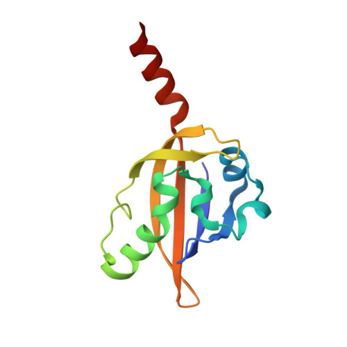
The FixL protein of Bradyrhizobium japonicum is a dimeric oxygen sensor responsible for initiating regulation of transcription of genes encoding proteins involved in nitrogen fixation and oxidative stress. It consists of an N-terminal heme-bound PAS domain, denoted bjFixLH, and a C-terminal histidine kinase domain whose enzymatic activity depends on the ligation state of the heme. To investigate the molecular basis for this dependence and the dynamics associated with conversion between ligated and unligated states, we have conducted time-resolved Laue diffraction studies of CO recombination in bjFixLH. Time-dependent difference Fourier maps from 1 micros to 10 ms after photolysis of the heme-CO bond show movement of the side chain of Leu236 and the H and I beta-strands into the ligand binding pocket formerly occupied by CO. Long-range conformational changes are evident in the protein, driven by relaxation of steric interactions between the bound ligand and amino acid side chains and/or changes in heme stereochemistry. These structural changes fully reverse as CO rebinds to the heme. Spectroscopic measurements of CO recombination kinetics in bjFixLH crystals relate the behavior of crystalline bjFixLH to solution and provide a framework for our time-resolved crystallographic experiments. Analysis of the time-dependent difference Fourier maps by singular value decomposition reveals that only one significant singular value accounts for the data. Thus only two structural states are present, the photolyzed and the CO-bound states. The first left singular vector represents the difference in density between these two states and shows features common to difference maps calculated from the static CO and deoxy states. The first right singular vector represents the time course of this difference density and agrees well with the CO recombination kinetics measured spectroscopically. We refine the structure of the photolyzed state present in the early-microsecond time range and find that it does not differ significantly in conformation from static, deoxy bjFixLH. Thus, structural relaxation from CO-bound to deoxy bjFixLH is complete in less than 1 micros.
Organizational Affiliation:
Department of Biochemistry and Molecular Biology, University of Chicago, Chicago, Illinois 60637, USA.