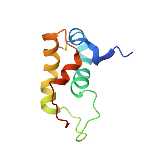
HdeB chaperone activity is coupled to its intrinsic dynamic properties.
Ding, J., Yang, C., Niu, X., Hu, Y., Jin, C.(2015) Sci Rep 5: 16856-16856
- PubMed: 26593705
- DOI: https://doi.org/10.1038/srep16856
- Primary Citation of Related Structures:
2MYJ - PubMed Abstract:
Enteric bacteria encounter extreme acidity when passing through hosts' stomach. Since the bacterial periplasmic space quickly equilibrates with outer environment, an efficient acid resistance mechanism is essential in preventing irreversible protein denaturation/aggregation and maintaining bacteria viability. HdeB, along with its homolog HdeA, was identified as a periplasmic acid-resistant chaperone. Both proteins exist as homodimers and share similar monomeric structures under neutral pH, while showing different dimeric packing interfaces. Previous investigations show that HdeA functions through an acid-induced dimer-to-monomer transition and partial unfolding at low pH (pH 2-3), resulting in exposure of hydrophobic surfaces that bind substrate proteins. In contrast, HdeB appears to have a much higher optimal activation pH (pH 4-5), under which condition the protein maintains a well-folded dimer and the mechanism for its chaperone activity remains elusive. Herein, we present an NMR study of HdeB to investigate its dynamic properties. Our results reveal that HdeB undergoes significant micro- to milli-second timescale conformational exchanges at neutral to near-neutral pH, under the later condition it exhibits optimal activity. The current study indicates that HdeB activation is coupled to its intrinsic dynamics instead of structural changes, and therefore its functional mechanism is apparently different from HdeA.
Organizational Affiliation:
College of Life Sciences, Peking University, Beijing 100871, China.