2MAS

PURINE NUCLEOSIDE HYDROLASE WITH A TRANSITION STATE INHIBITOR


Experimental Data Snapshot

  • Method: X-RAY DIFFRACTION
  • Resolution: 2.30 Å
  • R-Value Free: 0.234 
  • R-Value Work: 0.205 
  • R-Value Observed: 0.205 

wwPDB Validation   3D Report Full Report


This is version 1.3 of the entry. See complete history


Literature

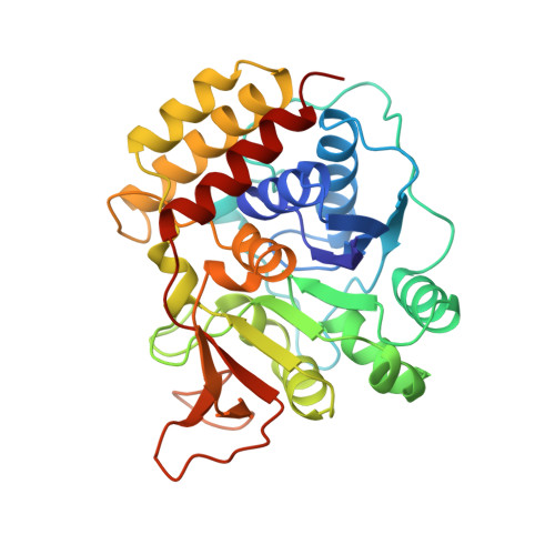
Trypanosomal nucleoside hydrolase. A novel mechanism from the structure with a transition-state inhibitor.

Degano, M.Almo, S.C.Sacchettini, J.C.Schramm, V.L.

(1998) Biochemistry 37: 6277-6285

  • DOI: https://doi.org/10.1021/bi973012e
  • Primary Citation of Related Structures:  
    2MAS

  • PubMed Abstract: 

    Nucleoside N-ribohydrolases are targets for disruption of purine salvage in the protozoan parasites. The structure of a trypanosomal N-ribohydrolase in complex with a transition-state inhibitor is reported at 2.3 A resolution. The nonspecific nucleoside hydrolase from Crithidia fasciculata cocrystallized with p-aminophenyliminoribitol reveals tightly bound Ca2+ as a catalytic site ligand. The complex with the transition-state inhibitor is characterized by (1) large protein conformational changes to create a hydrophobic leaving group site (2) C3'-exo geometry for the inhibitor, typical of a ribooxocarbenium ion (3) stabilization of the ribooxocarbenium analogue between the neighboring group 5'-hydroxyl and bidentate hydrogen bonds to Asn168; and (4) octacoordinate Ca2+ orients a catalytic site water and is liganded to two hydroxyls of the inhibitor. The mechanism is ribooxocarbenium stabilization with weak leaving group activation and is a departure from glucohydrolases which use paired carboxylates to achieve the transition state.


  • Organizational Affiliation

    Department of Biochemistry, Albert Einstein College of Medicine, Bronx, New York 10461, USA.


Macromolecules
Find similar proteins by:  (by identity cutoff)  |  3D Structure
Entity ID: 1
MoleculeChains Sequence LengthOrganismDetailsImage
INOSINE-URIDINE NUCLEOSIDE N-RIBOHYDROLASE
A, B, C, D
314Crithidia fasciculataMutation(s): 0 
Gene Names: IU-NH FROM C. FASCICULATA
EC: 3.2.2.1
UniProt
Find proteins for Q27546 (Crithidia fasciculata)
Explore Q27546 
Go to UniProtKB:  Q27546
Entity Groups  
Sequence Clusters30% Identity50% Identity70% Identity90% Identity95% Identity100% Identity
UniProt GroupQ27546
Sequence Annotations
Expand
  • Reference Sequence
Experimental Data & Validation

Experimental Data

  • Method: X-RAY DIFFRACTION
  • Resolution: 2.30 Å
  • R-Value Free: 0.234 
  • R-Value Work: 0.205 
  • R-Value Observed: 0.205 
  • Space Group: I 2 2 2
Unit Cell:
Length ( Å )Angle ( ˚ )
a = 120.63α = 90
b = 159.76β = 90
c = 206.38γ = 90
Software Package:
Software NamePurpose
X-PLORmodel building
X-PLORrefinement
SAINTdata reduction
SAINTdata scaling
X-PLORphasing

Structure Validation

View Full Validation Report


Entry History 

Deposition Data

Revision History  (Full details and data files)

  • Version 1.0: 1997-08-12
    Type: Initial release
  • Version 1.1: 2008-03-03
    Changes: Version format compliance
  • Version 1.2: 2011-07-13
    Changes: Version format compliance
  • Version 1.3: 2023-08-09
    Changes: Database references, Derived calculations, Other, Refinement description