High-definition NMR structure of PED/PEA-15 death effector domain reveals details of key polar side chain interactions.
Twomey, E.C., Wei, Y.(2012) Biochem Biophys Res Commun 424: 141-146
- PubMed: 22732408
- DOI: https://doi.org/10.1016/j.bbrc.2012.06.091
- Primary Citation of Related Structures:
2LS7 - PubMed Abstract:
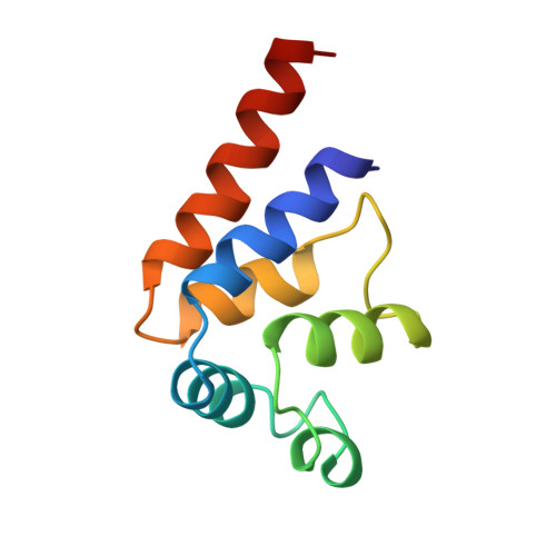
Death effector domain (DED) proteins constitute a subfamily of the large death domain superfamily that is primarily involved in apoptosis pathways. DED structures have characteristic side chain-side chain interactions among polar residues on the protein surface, forming a network of hydrogen bonds and salt bridges. The polar interaction network is functionally important in promoting protein-protein interactions by maintaining optimal side chain orientations. We have refined the solution DED structure of the PED/PEA-15 protein, a representative member of DED subfamily, using traditional NMR restraints with the addition of residual dipolar coupling (RDC) restraints from two independent alignment media, and employed the explicit solvent refinement protocol. The newly refined DED structure of PED/PEA-15 possesses higher structural quality as indicated by WHAT IF Z-scores, with most significant improvement in the backbone conformation normality quality factor. This higher quality DED structure of PED/PEA-15 leads to the identification of a number of key polar side chain interactions, which are not typically observed in NMR protein structures. The elucidation of polar side chain interactions is a key step towards the understanding of protein-protein interactions involving the death domain superfamily. The NMR structures with extensive details of protein structural features are thereby termed high-definition (HD) NMR structures.
Organizational Affiliation:
Department of Chemistry and Biochemistry, Seton Hall University, South Orange, NJ 07094-2646, USA.