2IV2

Reinterpretation of reduced form of formate dehydrogenase H from E. coli


Experimental Data Snapshot

  • Method: X-RAY DIFFRACTION
  • Resolution: 2.27 Å
  • R-Value Free: 0.226 
  • R-Value Work: 0.175 
  • R-Value Observed: 0.178 

wwPDB Validation   3D Report Full Report


This is version 1.3 of the entry. See complete history


Literature

Formate-Reduced E. Coli Formate Dehydrogenase H: The Reinterpretation of the Crystal Structure Suggests a New Reaction Mechanism.

Raaijmakers, H.C.A.Romao, M.J.

(2006) J Biol Inorg Chem 11: 849

  • DOI: https://doi.org/10.1007/s00775-006-0129-2
  • Primary Citation of Related Structures:  
    2IV2

  • PubMed Abstract: 

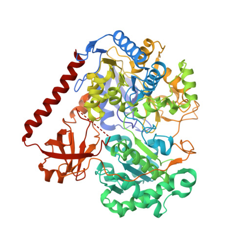
    Re-evaluation of the crystallographic data of the molybdenum-containing E. coli formate dehydrogenase H (Boyington et al. Science 275:1305-1308, 1997), reported in two redox states, reveals important structural differences for the formate-reduced form, with large implications for the reaction mechanism proposed in that work. We have re-refined the reduced structure with revised protocols and found substantial rearrangement in some parts of it. The original model is essentially correct but an important loop close to the molybdenum active site was mistraced, and, therefore, catalytic relevant residues were located in wrong positions. In particular selenocysteine-140, a ligand of molybdenum in the original work, and essential for catalysis, is no longer bound to the metal after reduction of the enzyme with formate. These results are incompatible with the originally proposed reaction mechanism. On the basis of our new interpretation, we have revised and proposed a new reaction mechanism, which reconciles the new X-ray model with previous biochemical and extended X-ray absorption fine structure data.


  • Organizational Affiliation

    REQUIMTE/CQFB, Departamento de Química, Faculdade de Ciências e Tecnologia, Universidade Nova de Lisboa, 2829-516, Monte de Caparica, Portugal.


Macromolecules
Find similar proteins by:  (by identity cutoff)  |  3D Structure
Entity ID: 1
MoleculeChains Sequence LengthOrganismDetailsImage
Formate dehydrogenase HA [auth X]715Escherichia coliMutation(s): 0 
Gene Names: fdhFb4079JW4040
EC: 1.17.1.9 (PDB Primary Data), 1.17.99.7 (PDB Primary Data)
UniProt
Find proteins for P07658 (Escherichia coli (strain K12))
Explore P07658 
Go to UniProtKB:  P07658
Entity Groups  
Sequence Clusters30% Identity50% Identity70% Identity90% Identity95% Identity100% Identity
UniProt GroupP07658
Sequence Annotations
Expand
  • Reference Sequence
Small Molecules
Ligands 5 Unique
IDChains Name / Formula / InChI Key2D Diagram3D Interactions
2MD
Query on 2MD

Download Ideal Coordinates CCD File 
C [auth X]GUANYLATE-O'-PHOSPHORIC ACID MONO-(2-AMINO-5,6-DIMERCAPTO-4-OXO-3,5,6,7,8A,9,10,10A-OCTAHYDRO-4H-8-OXA-1,3,9,10-TETRAAZA-ANTHRACEN-7-YLMETHYL) ESTER
C20 H28 N10 O13 P2 S2
YDYCAANTTOPADF-JKOCJXFLSA-N
MGD
Query on MGD

Download Ideal Coordinates CCD File 
D [auth X]2-AMINO-5,6-DIMERCAPTO-7-METHYL-3,7,8A,9-TETRAHYDRO-8-OXA-1,3,9,10-TETRAAZA-ANTHRACEN-4-ONE GUANOSINE DINUCLEOTIDE
C20 H26 N10 O13 P2 S2
VQAGYJCYOLHZDH-ILXWUORBSA-N
SF4
Query on SF4

Download Ideal Coordinates CCD File 
B [auth X]IRON/SULFUR CLUSTER
Fe4 S4
LJBDFODJNLIPKO-UHFFFAOYSA-N
MO
Query on MO

Download Ideal Coordinates CCD File 
E [auth X]MOLYBDENUM ATOM
Mo
ZOKXTWBITQBERF-UHFFFAOYSA-N
UNX
Query on UNX

Download Ideal Coordinates CCD File 
F [auth X]UNKNOWN ATOM OR ION
X
Experimental Data & Validation

Experimental Data

  • Method: X-RAY DIFFRACTION
  • Resolution: 2.27 Å
  • R-Value Free: 0.226 
  • R-Value Work: 0.175 
  • R-Value Observed: 0.178 
  • Space Group: P 41 21 2
Unit Cell:
Length ( Å )Angle ( ˚ )
a = 146.4α = 90
b = 146.4β = 90
c = 81.27γ = 90
Software Package:
Software NamePurpose
REFMACrefinement

Structure Validation

View Full Validation Report


Entry History 

Deposition Data

Revision History  (Full details and data files)

  • Version 1.0: 2006-06-12
    Type: Initial release
  • Version 1.1: 2014-02-19
    Changes: Atomic model, Derived calculations, Non-polymer description, Other, Structure summary, Version format compliance
  • Version 1.2: 2018-12-19
    Changes: Data collection, Database references, Source and taxonomy, Structure summary
  • Version 1.3: 2023-12-13
    Changes: Data collection, Database references, Derived calculations, Refinement description