2I6E

Crystal structure of protein DR0370 from Deinococcus radiodurans, Pfam DUF178


Experimental Data Snapshot

  • Method: X-RAY DIFFRACTION
  • Resolution: 2.50 Å
  • R-Value Free: 0.235 
  • R-Value Work: 0.202 
  • R-Value Observed: 0.202 

wwPDB Validation   3D Report Full Report


This is version 1.3 of the entry. See complete history


Literature

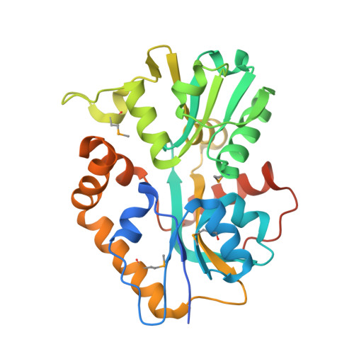
X-ray structures of two proteins belonging to Pfam DUF178 revealed unexpected structural similarity to the DUF191 Pfam family.

Tyagi, R.Burley, S.K.Swaminathan, S.

(2007) BMC Struct Biol 7: 62-62

  • DOI: https://doi.org/10.1186/1472-6807-7-62
  • Primary Citation of Related Structures:  
    2I6E

  • PubMed Abstract: 

    Pfam is a comprehensive collection of protein domains and families, with a range of well-established information including genome annotation. Pfam has two large series of functionally uncharacterized families, known as Domains of Unknown Function (DUFs) and Uncharacterized Protein Families (UPFs). Crystal structures of two proteins from Deinococcus radiodurans and Streptomyces coelicolor belonging to Pfam protein family DUF178 (ID: PF02621) have been determined using Selenium-Single-wavelength Anomalous Dispersion (Se-SAD). Based on the structure, we have identified the putative function for this family of protein. Unexpectedly, we found that DUF178 Pfam is remarkably similar to Pfam family DUF191 suggesting that the sequence-based classification alone may not be sufficient to classify proteins into Pfam families.


  • Organizational Affiliation

    Biology Department, Brookhaven National Laboratory, Upton, New York 11973, USA. swami@bnl.gov


Macromolecules
Find similar proteins by:  (by identity cutoff)  |  3D Structure
Entity ID: 1
MoleculeChains Sequence LengthOrganismDetailsImage
Hypothetical protein
A, B, C, D, E
A, B, C, D, E, F, G, H
301Deinococcus radioduransMutation(s): 4 
Gene Names: DR_0370
UniProt
Find proteins for Q9RXE3 (Deinococcus radiodurans (strain ATCC 13939 / DSM 20539 / JCM 16871 / CCUG 27074 / LMG 4051 / NBRC 15346 / NCIMB 9279 / VKM B-1422 / R1))
Explore Q9RXE3 
Go to UniProtKB:  Q9RXE3
Entity Groups  
Sequence Clusters30% Identity50% Identity70% Identity90% Identity95% Identity100% Identity
UniProt GroupQ9RXE3
Sequence Annotations
Expand
  • Reference Sequence
Small Molecules
Ligands 1 Unique
IDChains Name / Formula / InChI Key2D Diagram3D Interactions
SO4
Query on SO4

Download Ideal Coordinates CCD File 
I [auth A]
J [auth A]
K [auth B]
L [auth B]
M [auth B]
I [auth A],
J [auth A],
K [auth B],
L [auth B],
M [auth B],
N [auth C],
O [auth C],
P [auth D],
Q [auth D],
R [auth E],
S [auth E],
T [auth F],
U [auth F],
V [auth F],
W [auth G],
X [auth G],
Y [auth G],
Z [auth H]
SULFATE ION
O4 S
QAOWNCQODCNURD-UHFFFAOYSA-L
Modified Residues  1 Unique
IDChains TypeFormula2D DiagramParent
MSE
Query on MSE
A, B, C, D, E
A, B, C, D, E, F, G, H
L-PEPTIDE LINKINGC5 H11 N O2 SeMET
Experimental Data & Validation

Experimental Data

  • Method: X-RAY DIFFRACTION
  • Resolution: 2.50 Å
  • R-Value Free: 0.235 
  • R-Value Work: 0.202 
  • R-Value Observed: 0.202 
  • Space Group: P 1 21 1
Unit Cell:
Length ( Å )Angle ( ˚ )
a = 75.31α = 90
b = 139.37β = 92.79
c = 153.615γ = 90
Software Package:
Software NamePurpose
CNSrefinement
CBASSdata collection
HKL-2000data reduction
HKL-2000data scaling
SHELXDphasing
SHARPphasing

Structure Validation

View Full Validation Report


Entry History 

Deposition Data

Revision History  (Full details and data files)

  • Version 1.0: 2006-09-05
    Type: Initial release
  • Version 1.1: 2007-10-11
    Changes: Version format compliance
  • Version 1.2: 2011-07-13
    Changes: Derived calculations, Version format compliance
  • Version 1.3: 2021-02-03
    Changes: Database references, Derived calculations, Structure summary