


Crystal structure of a probable acetyltransferase
Eswaramoorthy, S., Swaminathan, S.To be published.
Experimental Data Snapshot
wwPDB Validation 3D Report Full Report

Entity ID: 1 | |||||
|---|---|---|---|---|---|
| Molecule | Chains | Sequence Length | Organism | Details | Image |
| Probable acetyltransferase | A, B, C [auth D], D [auth E] | 153 | Pseudomonas aeruginosa PAO1 | Mutation(s): 4 | 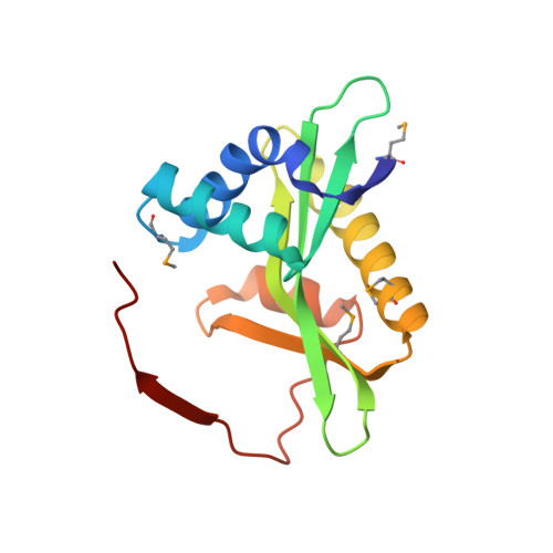 |
UniProt | |||||
Find proteins for Q9HX01 (Pseudomonas aeruginosa (strain ATCC 15692 / DSM 22644 / CIP 104116 / JCM 14847 / LMG 12228 / 1C / PRS 101 / PAO1)) Explore Q9HX01 Go to UniProtKB: Q9HX01 | |||||
Entity Groups | |||||
| Sequence Clusters | 30% Identity50% Identity70% Identity90% Identity95% Identity100% Identity | ||||
| UniProt Group | Q9HX01 | ||||
Sequence AnnotationsExpand | |||||
| |||||
| Modified Residues 1 Unique | |||||
|---|---|---|---|---|---|
| ID | Chains | Type | Formula | 2D Diagram | Parent |
| MSE Query on MSE | A, B, C [auth D], D [auth E] | L-PEPTIDE LINKING | C5 H11 N O2 Se |  | MET |
| Length ( Å ) | Angle ( ˚ ) |
|---|---|
| a = 115.1 | α = 90 |
| b = 115.1 | β = 90 |
| c = 66.85 | γ = 90 |
| Software Name | Purpose |
|---|---|
| CBASS | data collection |
| SCALEPACK | data scaling |
| SOLVE | phasing |
| CNS | refinement |


