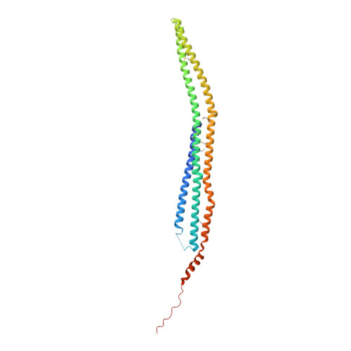
2EFK

Crystal structure of the EFC domain of Cdc42-interacting protein 4


Experimental Data Snapshot

  • Method: X-RAY DIFFRACTION
  • Resolution: 2.30 Å
  • R-Value Free: 0.279 
  • R-Value Work: 0.228 
  • R-Value Observed: 0.228 

wwPDB Validation   3D Report Full Report


This is version 1.2 of the entry. See complete history


Literature

Curved EFC/F-BAR-Domain Dimers Are Joined End to End into a Filament for Membrane Invagination in Endocytosis

Shimada, A.Niwa, H.Tsujita, K.Suetsugu, S.Nitta, K.Hanawa-Suetsugu, K.Akasaka, R.Nishino, Y.Toyama, M.Chen, L.Liu, Z.-J.Wang, B.-C.Yamamoto, M.Terada, T.Miyazawa, A.Tanaka, A.Sugano, S.Shirouzu, M.Nagayama, K.Takenawa, T.Yokoyama, S.

(2007) Cell 129: 761-772

  • DOI: https://doi.org/10.1016/j.cell.2007.03.040
  • Primary Citation of Related Structures:  
    2EFK, 2EFL

  • PubMed Abstract: 

    Pombe Cdc15 homology (PCH) proteins play an important role in a variety of actin-based processes, including clathrin-mediated endocytosis (CME). The defining feature of the PCH proteins is an evolutionarily conserved EFC/F-BAR domain for membrane association and tubulation. In the present study, we solved the crystal structures of the EFC domains of human FBP17 and CIP4. The structures revealed a gently curved helical-bundle dimer of approximately 220 A in length, which forms filaments through end-to-end interactions in the crystals. The curved EFC dimer fits a tubular membrane with an approximately 600 A diameter. We subsequently proposed a model in which the curved EFC filament drives tubulation. In fact, striation of tubular membranes was observed by phase-contrast cryo-transmission electron microscopy, and mutations that impaired filament formation also impaired membrane tubulation and cell membrane invagination. Furthermore, FBP17 is recruited to clathrin-coated pits in the late stage of CME, indicating its physiological role.


  • Organizational Affiliation

    RIKEN SPring-8 Center, Harima Institute, 1-1-1 Kouto, Sayo, Hyogo 679-5148, Japan.


Macromolecules
Find similar proteins by:  (by identity cutoff)  |  3D Structure
Entity ID: 1
MoleculeChains Sequence LengthOrganismDetailsImage
Cdc42-interacting protein 4301Homo sapiensMutation(s): 7 
Membrane Entity: Yes 
UniProt & NIH Common Fund Data Resources
Find proteins for Q15642 (Homo sapiens)
Explore Q15642 
Go to UniProtKB:  Q15642
PHAROS:  Q15642
GTEx:  ENSG00000125733 
Entity Groups  
Sequence Clusters30% Identity50% Identity70% Identity90% Identity95% Identity100% Identity
UniProt GroupQ15642
Sequence Annotations
Expand
  • Reference Sequence
Small Molecules
Modified Residues  1 Unique
IDChains TypeFormula2D DiagramParent
MSE
Query on MSE
A
L-PEPTIDE LINKINGC5 H11 N O2 SeMET
Experimental Data & Validation

Experimental Data

  • Method: X-RAY DIFFRACTION
  • Resolution: 2.30 Å
  • R-Value Free: 0.279 
  • R-Value Work: 0.228 
  • R-Value Observed: 0.228 
  • Space Group: C 1 2 1
Unit Cell:
Length ( Å )Angle ( ˚ )
a = 94.817α = 90
b = 70.41β = 107.2
c = 65.679γ = 90
Software Package:
Software NamePurpose
CNSrefinement
HKL-2000data collection
HKL-2000data reduction
HKL-2000data scaling
CNSphasing

Structure Validation

View Full Validation Report


Entry History 

Revision History  (Full details and data files)

  • Version 1.0: 2007-05-29
    Type: Initial release
  • Version 1.1: 2008-04-30
    Changes: Version format compliance
  • Version 1.2: 2011-07-13
    Changes: Derived calculations, Version format compliance