NMR structure of mussel mytilin, and antiviral-antibacterial activities of derived synthetic peptides.
Roch, P., Yang, Y., Toubiana, M., Aumelas, A.(2008) Dev Comp Immunol 32: 227-238
- PubMed: 17628674
- DOI: https://doi.org/10.1016/j.dci.2007.05.006
- Primary Citation of Related Structures:
2EEM - PubMed Abstract:
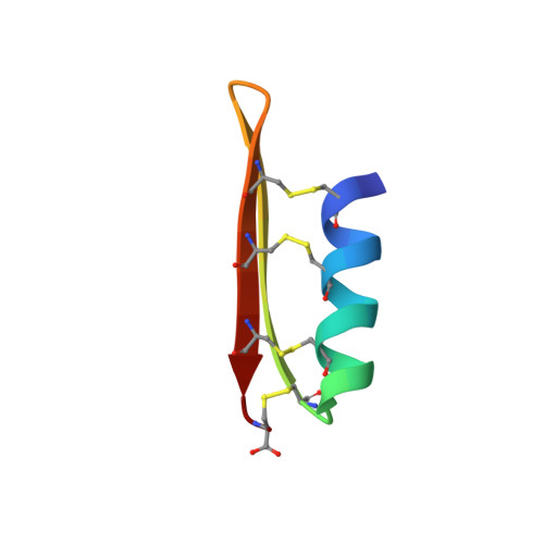
Mytilin is a 34-residue antibacterial peptide from the mussel Mytilus galloprovincialis, which in addition possesses in vitro antiviral activity. The three-dimensional solution structure of the synthetic mytilin was established by using 1H NMR and consists of the common cysteine-stabilized alphabeta motif close to the one observed in the mussel defensin MGD-1. Mytilin is characterized by 8 cysteines engaged in four disulfide bonds (2-27, 6-29, 10-31, and 15-34) only involving the beta-strand II. Hydrophilic and hydrophobic areas of mytilin account for 63% and 37%, respectively, a ratio very close to that of MGD-1 (64% and 36%). One linear and three cyclic fragments were designed from the interstrand loop sequence known to retain the biological activities in MGD-1. Only the fragment of 10 amino acids (C10C) constrained by two disulfide bonds in a stable beta-hairpin structure was able to inhibit the mortality of Palaemon serratus shrimp injected with white spot syndrome virus (WSSV). Fifty percent inhibition was obtained by in vitro pre-incubation of WSSV with 45 microM of C10C compared with 7 microM for mytilin. Interaction between the fragment and the virus occurred very rapidly as 40% survival was recorded after only 1 min of pre-incubation. In addition, C10C was capable of inhibiting in vitro growth of Vibrio splendidus LGP32 (MIC 125 microM), Vibrio anguillarum (MIC 2mM), Micrococcus lysodeikticus and Escherichia coli (MIC 1mM). Destroying the cysteine-stabilized alphabeta structure or shortening the C10C fragment to the C6C fragment with only one disulfide bond resulted in loss of both antiviral and antibacterial activities. Increasing the positive net charge did not enforce the antibacterial activity and completely suppressed the antiviral one. The C10C-designed peptide from mytilin appeared comparable in composition and structure with protegrin, tachyplesin and polyphemusin.
Organizational Affiliation:
CNRS UMR5119-IFREMER-Université Montpellier 2, Ecosystèmes Lagunaires, cc093, place E. Bataillon, F-34095 Montpellier, France. philippe.roch@univ-montp2.fr