2D04

Crystal structure of neoculin, a sweet protein with taste-modifying activity.


Experimental Data Snapshot

  • Method: X-RAY DIFFRACTION
  • Resolution: 2.76 Å
  • R-Value Free: 0.267 
  • R-Value Work: 0.245 
  • R-Value Observed: 0.245 

wwPDB Validation   3D Report Full Report


Ligand Structure Quality Assessment 


This is version 2.1 of the entry. See complete history


Literature

Crystal structure of neoculin: insights into its sweetness and taste-modifying activity

Shimizu-Ibuka, A.Morita, Y.Terada, T.Asakura, T.Nakajima, K.Iwata, S.Misaka, T.Sorimachi, H.Arai, S.Abe, K.

(2006) J Mol Biol 359: 148-158

  • DOI: https://doi.org/10.1016/j.jmb.2006.03.030
  • Primary Citation of Related Structures:  
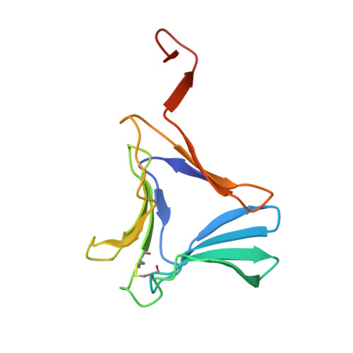
    2D04

  • PubMed Abstract: 

    Although the majority of sweet compounds are of low molecular mass, several proteins are known to elicit sweet taste responses in humans. The fruit of Curculigo latifolia contains a heterodimeric protein, neoculin, which has both sweetness and a taste-modifying activity that converts sourness to sweetness. Here, we report the crystal structure of neoculin at 2.76A resolution. This is the first well-defined tertiary structure of a taste-modifying protein of this kind. The overall structure is quite similar to those of monocot mannose-binding lectins. However, crucial topological differences are observed in the C-terminal regions of both subunits. In both subunits of neoculin, the C-terminal tails turn up to form loops fixed by inter-subunit disulfide bonds that are not observed in the lectins. Indeed, the corresponding regions of the lectins stretch straight over the surface of another subunit. Such a C-terminal structural feature as is observed in neoculin results in a decrease in subunit-subunit interactions. Moreover, distribution of electrostatic potential on the surface of neoculin is unique and significantly different from those of the lectins, particularly in the basic subunit (NBS). We have found that there is a large cluster composed of six basic residues on the surface of NBS, and speculate that it might be involved in the elicitation of sweetness and/or taste-modifying activity of neoculin. Molecular dynamics simulation based on the crystallography results suggests that neoculin may adopt a widely "open" conformation at acidic pH, while unprotonated neoculin at neutral pH is in a "closed" conformation. Based on these simulations and the generation of a docking model between neoculin and the sweet-taste receptor, T1R2-T1R3, we propose the hypothesis that neoculin is in dynamic equilibrium between open and closed states, and that the addition of an acid shifts the equilibrium to the open state, allowing ligand-receptor interaction.


  • Organizational Affiliation

    Department of Applied Biological Chemistry, Graduate School of Agricultural and Life Sciences, The University of Tokyo, Bunkyo-ku, Japan.


Macromolecules
Find similar proteins by:  (by identity cutoff)  |  3D Structure
Entity ID: 1
MoleculeChains Sequence LengthOrganismDetailsImage
neoculin acidic subunit
A, C, E, G
113Molineria latifoliaMutation(s): 0 
UniProt
Find proteins for Q6F495 (Molineria latifolia)
Explore Q6F495 
Go to UniProtKB:  Q6F495
Entity Groups  
Sequence Clusters30% Identity50% Identity70% Identity90% Identity95% Identity100% Identity
UniProt GroupQ6F495
Sequence Annotations
Expand
  • Reference Sequence
Find similar proteins by:  (by identity cutoff)  |  3D Structure
Entity ID: 2
MoleculeChains Sequence LengthOrganismDetailsImage
Curculin
B, D, F, H
114Molineria latifoliaMutation(s): 0 
UniProt
Find proteins for P19667 (Molineria latifolia)
Explore P19667 
Go to UniProtKB:  P19667
Entity Groups  
Sequence Clusters30% Identity50% Identity70% Identity90% Identity95% Identity100% Identity
UniProt GroupP19667
Sequence Annotations
Expand
  • Reference Sequence
Oligosaccharides

Help

Entity ID: 3
MoleculeChains Length2D Diagram Glycosylation3D Interactions
beta-D-mannopyranose-(1-4)-2-acetamido-2-deoxy-beta-D-glucopyranose-(1-4)-[beta-L-fucopyranose-(1-3)]2-acetamido-2-deoxy-beta-D-glucopyranose
I
4N-Glycosylation
Glycosylation Resources
GlyTouCan:  G77577HJ
GlyCosmos:  G77577HJ
GlyGen:  G77577HJ
Small Molecules
Ligands 1 Unique
IDChains Name / Formula / InChI Key2D Diagram3D Interactions
NAG
Query on NAG

Download Ideal Coordinates CCD File 
J [auth A]2-acetamido-2-deoxy-beta-D-glucopyranose
C8 H15 N O6
OVRNDRQMDRJTHS-FMDGEEDCSA-N
Experimental Data & Validation

Experimental Data

  • Method: X-RAY DIFFRACTION
  • Resolution: 2.76 Å
  • R-Value Free: 0.267 
  • R-Value Work: 0.245 
  • R-Value Observed: 0.245 
  • Space Group: P 21 21 21
Unit Cell:
Length ( Å )Angle ( ˚ )
a = 48.009α = 90
b = 101.089β = 90
c = 271.572γ = 90
Software Package:
Software NamePurpose
CNSrefinement
HKL-2000data reduction
SCALEPACKdata scaling
PHASERphasing

Structure Validation

View Full Validation Report


Ligand Structure Quality Assessment 


Entry History 

Deposition Data

Revision History  (Full details and data files)

  • Version 1.0: 2006-06-20
    Type: Initial release
  • Version 1.1: 2008-04-30
    Changes: Version format compliance
  • Version 1.2: 2011-07-13
    Changes: Non-polymer description, Version format compliance
  • Version 2.0: 2020-07-29
    Type: Remediation
    Reason: Carbohydrate remediation
    Changes: Advisory, Atomic model, Data collection, Derived calculations, Structure summary
  • Version 2.1: 2023-10-25
    Changes: Advisory, Data collection, Database references, Refinement description, Structure summary