2C1L

Structure of the BfiI restriction endonuclease


Experimental Data Snapshot

  • Method: X-RAY DIFFRACTION
  • Resolution: 1.90 Å
  • R-Value Free: 0.217 
  • R-Value Work: 0.188 
  • R-Value Observed: 0.188 

wwPDB Validation   3D Report Full Report


Ligand Structure Quality Assessment 


This is version 1.2 of the entry. See complete history


Literature

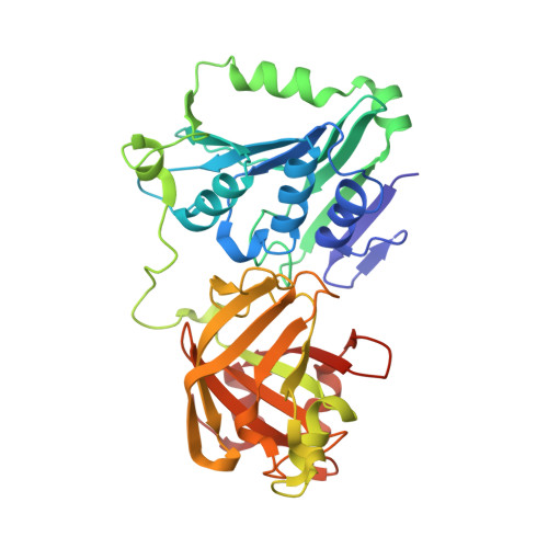
Structure of the Metal-Independent Restriction Enzyme Bfii Reveals Fusion of a Specific DNA-Binding Domain with a Nonspecific Nuclease.

Grazulis, S.Manakova, E.Roessle, M.Bochtler, M.Tamulaitiene, G.Huber, R.Siksnys, V.

(2005) Proc Natl Acad Sci U S A 102: 15797

  • DOI: https://doi.org/10.1073/pnas.0507949102
  • Primary Citation of Related Structures:  
    2C1L

  • PubMed Abstract: 

    Among all restriction endonucleases known to date, BfiI is unique in cleaving DNA in the absence of metal ions. BfiI represents a different evolutionary lineage of restriction enzymes, as shown by its crystal structure at 1.9-A resolution. The protein consists of two structural domains. The N-terminal catalytic domain is similar to Nuc, an EDTA-resistant nuclease from the phospholipase D superfamily. The C-terminal DNA-binding domain of BfiI exhibits a beta-barrel-like structure very similar to the effector DNA-binding domain of the Mg(2+)-dependent restriction enzyme EcoRII and to the B3-like DNA-binding domain of plant transcription factors. BfiI presumably evolved through domain fusion of a DNA-recognition element to a nonspecific nuclease akin to Nuc and elaborated a mechanism to limit DNA cleavage to a single double-strand break near the specific recognition sequence. The crystal structure suggests that the interdomain linker may act as an autoinhibitor controlling BfiI catalytic activity in the absence of a specific DNA sequence. A psi-blast search identified a BfiI homologue in a Mesorhizobium sp. BNC1 bacteria strain, a plant symbiont isolated from an EDTA-rich environment.


  • Organizational Affiliation

    Laboratory of Protein-DNA Interaction, Institute of Biotechnology, Graiciuno 8, LT-02241 Vilnius, Lithuania. grazulis@ibt.lt


Macromolecules
Find similar proteins by:  (by identity cutoff)  |  3D Structure
Entity ID: 1
MoleculeChains Sequence LengthOrganismDetailsImage
RESTRICTION ENDONUCLEASE
A, B
358Cytobacillus firmusMutation(s): 0 
EC: 3.1.21.4
UniProt
Find proteins for Q9F4C9 (Cytobacillus firmus)
Explore Q9F4C9 
Go to UniProtKB:  Q9F4C9
Entity Groups  
Sequence Clusters30% Identity50% Identity70% Identity90% Identity95% Identity100% Identity
UniProt GroupQ9F4C9
Sequence Annotations
Expand
  • Reference Sequence
Small Molecules
Ligands 7 Unique
IDChains Name / Formula / InChI Key2D Diagram3D Interactions
MES
Query on MES

Download Ideal Coordinates CCD File 
L [auth B],
M [auth B]
2-(N-MORPHOLINO)-ETHANESULFONIC ACID
C6 H13 N O4 S
SXGZJKUKBWWHRA-UHFFFAOYSA-N
TAR
Query on TAR

Download Ideal Coordinates CCD File 
I [auth A]D(-)-TARTARIC ACID
C4 H6 O6
FEWJPZIEWOKRBE-LWMBPPNESA-N
SRT
Query on SRT

Download Ideal Coordinates CCD File 
H [auth A],
N [auth B]
S,R MESO-TARTARIC ACID
C4 H6 O6
FEWJPZIEWOKRBE-XIXRPRMCSA-N
TLA
Query on TLA

Download Ideal Coordinates CCD File 
G [auth A],
O [auth B]
L(+)-TARTARIC ACID
C4 H6 O6
FEWJPZIEWOKRBE-JCYAYHJZSA-N
TRS
Query on TRS

Download Ideal Coordinates CCD File 
F [auth A]2-AMINO-2-HYDROXYMETHYL-PROPANE-1,3-DIOL
C4 H12 N O3
LENZDBCJOHFCAS-UHFFFAOYSA-O
GOL
Query on GOL

Download Ideal Coordinates CCD File 
C [auth A],
D [auth A],
E [auth A],
K [auth B]
GLYCEROL
C3 H8 O3
PEDCQBHIVMGVHV-UHFFFAOYSA-N
BCT
Query on BCT

Download Ideal Coordinates CCD File 
J [auth A],
P [auth B]
BICARBONATE ION
C H O3
BVKZGUZCCUSVTD-UHFFFAOYSA-M
Experimental Data & Validation

Experimental Data

  • Method: X-RAY DIFFRACTION
  • Resolution: 1.90 Å
  • R-Value Free: 0.217 
  • R-Value Work: 0.188 
  • R-Value Observed: 0.188 
  • Space Group: I 4
Unit Cell:
Length ( Å )Angle ( ˚ )
a = 138.925α = 90
b = 138.925β = 90
c = 94.135γ = 90
Software Package:
Software NamePurpose
MOSFLMdata reduction
SCALAdata scaling
MLPHAREphasing
DMphasing
CNSrefinement

Structure Validation

View Full Validation Report


Ligand Structure Quality Assessment 


Entry History 

Deposition Data

Revision History  (Full details and data files)

  • Version 1.0: 2005-10-07
    Type: Initial release
  • Version 1.1: 2011-05-08
    Changes: Version format compliance
  • Version 1.2: 2011-07-13
    Changes: Version format compliance