2BHY

Crystal structure of Deinococcus radiodurans maltooligosyltrehalose trehalohydrolase in complex with trehalose


Experimental Data Snapshot

  • Method: X-RAY DIFFRACTION
  • Resolution: 1.50 Å
  • R-Value Free: 0.152 
  • R-Value Work: 0.128 
  • R-Value Observed: 0.129 

wwPDB Validation   3D Report Full Report


Ligand Structure Quality Assessment 


This is version 2.0 of the entry. See complete history


Literature

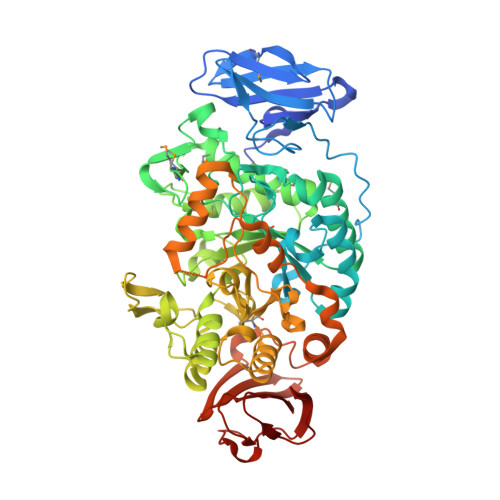
Crystal Structure of Maltooligosyltrehalose Trehalohydrolase from Deinococcus Radiodurans in Complex with Disaccharides

Timmins, J.Leiros, H.-K.S.Leonard, G.Leiros, I.Mcsweeney, S.

(2005) J Mol Biol 347: 949

  • DOI: https://doi.org/10.1016/j.jmb.2005.02.011
  • Primary Citation of Related Structures:  
    2BHU, 2BHY, 2BHZ

  • PubMed Abstract: 

    Trehalose (alpha-D-glucopyranosyl-1,1-alpha-D-glucopyranose) is a non-reducing diglucoside found in various organisms that serves as a carbohydrate reserve and as an agent that protects against a variety of physical and chemical stresses. Deinococcus radiodurans possesses an alternative biosynthesis pathway for the synthesis of trehalose from maltooligosaccharides. This reaction is mediated by two enzymes: maltooligosyltrehalose synthase (MTSase) and maltooligosyltrehalose trehalohydrolase (MTHase). Here, we present the 1.1A resolution crystal structure of MTHase. It consists of three major domains: two beta-sheet domains and a conserved glycosidase (beta/alpha)8 barrel catalytic domain. Three subdomains consisting of short insertions were identified within the catalytic domain. Subsequently, structures of MTHase in complex with maltose and trehalose were obtained at 1.2 A and 1.5 A resolution, respectively. These structures reveal the importance of the three inserted subdomains in providing the key residues required for substrate recognition. Trehalose is recognised specifically in the +1 and +2 binding subsites by an extensive hydrogen-bonding network and a strong hydrophobic stacking interaction in between two aromatic residues. Moreover, upon binding to maltose, which mimics the substrate sugar chain, a major concerted conformational change traps the sugar chain in the active site. The presence of magnesium in the active site of the MTHase-maltose complex suggests that MTHase activity may be regulated by divalent cations.


  • Organizational Affiliation

    Macromolecular Crystallography Group, European Synchrotron Radiation Facility, B.P. 220, 6 rue Jules Horowitz, F-38043 Grenoble Cedex, France.


Macromolecules
Find similar proteins by:  (by identity cutoff)  |  3D Structure
Entity ID: 1
MoleculeChains Sequence LengthOrganismDetailsImage
MALTOOLIGOSYLTREHALOSE TREHALOHYDROLASE602Deinococcus radiodurans R1 = ATCC 13939 = DSM 20539Mutation(s): 0 
EC: 3.2.1.1
UniProt
Find proteins for Q9RX51 (Deinococcus radiodurans (strain ATCC 13939 / DSM 20539 / JCM 16871 / CCUG 27074 / LMG 4051 / NBRC 15346 / NCIMB 9279 / VKM B-1422 / R1))
Explore Q9RX51 
Go to UniProtKB:  Q9RX51
Entity Groups  
Sequence Clusters30% Identity50% Identity70% Identity90% Identity95% Identity100% Identity
UniProt GroupQ9RX51
Sequence Annotations
Expand
  • Reference Sequence
Oligosaccharides

Help

Entity ID: 2
MoleculeChains Length2D Diagram Glycosylation3D Interactions
alpha-D-glucopyranose-(1-1)-alpha-D-glucopyranose
B, C, D, E, F
B, C, D, E, F, G, H
2N/A
Glycosylation Resources
GlyTouCan:  G92130SN
GlyCosmos:  G92130SN
Small Molecules
Modified Residues  1 Unique
IDChains TypeFormula2D DiagramParent
MSE
Query on MSE
A
L-PEPTIDE LINKINGC5 H11 N O2 SeMET
Experimental Data & Validation

Experimental Data

  • Method: X-RAY DIFFRACTION
  • Resolution: 1.50 Å
  • R-Value Free: 0.152 
  • R-Value Work: 0.128 
  • R-Value Observed: 0.129 
  • Space Group: P 21 21 21
Unit Cell:
Length ( Å )Angle ( ˚ )
a = 60.029α = 90
b = 66.556β = 90
c = 153.141γ = 90
Software Package:
Software NamePurpose
REFMACrefinement
MOSFLMdata reduction
SCALAdata scaling

Structure Validation

View Full Validation Report


Ligand Structure Quality Assessment 


Entry History 

Deposition Data

Revision History  (Full details and data files)

  • Version 1.0: 2005-03-31
    Type: Initial release
  • Version 1.1: 2011-05-08
    Changes: Version format compliance
  • Version 1.2: 2011-07-13
    Changes: Version format compliance
  • Version 2.0: 2020-07-29
    Type: Remediation
    Reason: Carbohydrate remediation
    Changes: Advisory, Atomic model, Data collection, Derived calculations, Non-polymer description, Other, Structure summary