2APH

Crystal structure of human PGRP-IalphaC in complex with muramyl pentapeptide


Experimental Data Snapshot

  • Method: X-RAY DIFFRACTION
  • Resolution: 2.10 Å
  • R-Value Free: 0.267 
  • R-Value Work: 0.227 
  • R-Value Observed: 0.227 

wwPDB Validation   3D Report Full Report


Ligand Structure Quality Assessment 


This is version 3.0 of the entry. See complete history


Literature

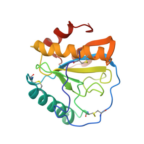
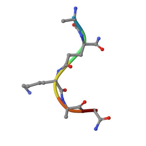
Crystal structure of human peptidoglycan recognition protein I alpha bound to a muramyl pentapeptide from Gram-positive bacteria.

Guan, R.Brown, P.H.Swaminathan, C.P.Roychowdhury, A.Boons, G.J.Mariuzza, R.A.

(2006) Protein Sci 15: 1199-1206

  • DOI: https://doi.org/10.1110/ps.062077606
  • Primary Citation of Related Structures:  
    2APH

  • PubMed Abstract: 

    Peptidoglycan recognition proteins (PGRPs) are pattern recognition receptors of the innate immune system that bind bacterial peptidoglycans (PGNs). We determined the crystal structure, to 2.1 A resolution, of the C-terminal PGN-binding domain of human PGRP-I alpha in complex with a muramyl pentapeptide (MPP) from Gram-positive bacteria containing a complete peptide stem (L-Ala-D-isoGln-L-Lys-D-Ala-D-Ala). The structure reveals important features not observed previously in the complex between PGRP-I alpha and a muramyl tripeptide lacking D-Ala at stem positions 4 and 5. Most notable are ligand-induced structural rearrangements in the PGN-binding site that are essential for entry of the C-terminal portion of the peptide stem and for locking MPP in the binding groove. We propose that similar structural rearrangements to accommodate the PGN stem likely characterize many PGRPs, both mammalian and insect.


  • Organizational Affiliation

    Center for Advanced Research in Biotechnology, W.M. Keck Laboratory for Structural Biology, University of Maryland Biotechnology Institute, Rockville, Maryland 20850, USA.


Macromolecules
Find similar proteins by:  (by identity cutoff)  |  3D Structure
Entity ID: 1
MoleculeChains Sequence LengthOrganismDetailsImage
Peptidoglycan recognition protein I-alpha
A, B
165Homo sapiensMutation(s): 0 
Gene Names: PGLYRP3PGRPIA
UniProt & NIH Common Fund Data Resources
Find proteins for Q96LB9 (Homo sapiens)
Explore Q96LB9 
Go to UniProtKB:  Q96LB9
PHAROS:  Q96LB9
GTEx:  ENSG00000159527 
Entity Groups  
Sequence Clusters30% Identity50% Identity70% Identity90% Identity95% Identity100% Identity
UniProt GroupQ96LB9
Sequence Annotations
Expand
  • Reference Sequence

Find similar proteins by:  Sequence   |   3D Structure  

Entity ID: 2
MoleculeChains Sequence LengthOrganismDetailsImage
muramyl pentapeptide
C, D
6synthetic constructMutation(s): 0 
Sequence Annotations
Expand
  • Reference Sequence
Experimental Data & Validation

Experimental Data

  • Method: X-RAY DIFFRACTION
  • Resolution: 2.10 Å
  • R-Value Free: 0.267 
  • R-Value Work: 0.227 
  • R-Value Observed: 0.227 
  • Space Group: P 1 21 1
Unit Cell:
Length ( Å )Angle ( ˚ )
a = 37.86α = 90
b = 58.63β = 93.65
c = 70.4γ = 90
Software Package:
Software NamePurpose
CNSrefinement
CrystalCleardata reduction
CrystalCleardata scaling
AMoREphasing

Structure Validation

View Full Validation Report


Ligand Structure Quality Assessment 


Entry History 

Deposition Data

Revision History  (Full details and data files)

  • Version 1.0: 2006-06-27
    Type: Initial release
  • Version 1.1: 2008-04-30
    Changes: Version format compliance
  • Version 1.2: 2011-07-13
    Changes: Derived calculations, Version format compliance
  • Version 1.3: 2018-04-04
    Changes: Advisory, Data collection
  • Version 1.4: 2020-07-29
    Type: Remediation
    Reason: Carbohydrate remediation
    Changes: Advisory, Data collection, Derived calculations, Structure summary
  • Version 1.5: 2023-08-23
    Changes: Data collection, Database references, Refinement description, Structure summary
  • Version 2.0: 2023-11-15
    Changes: Atomic model, Data collection, Derived calculations
  • Version 2.1: 2023-11-29
    Changes: Database references
  • Version 3.0: 2024-02-28
    Changes: Advisory, Atomic model, Data collection, Database references, Derived calculations, Polymer sequence, Source and taxonomy, Structure summary