232D

THE HIGH RESOLUTION CRYSTAL STRUCTURE OF THE DNA DECAMER D(AGGCATGCCT)

  • Classification: DNA
  • Mutation(s): No 

  • Deposited: 1995-09-05 Released: 1996-03-22 
  • Deposition Author(s): Nunn, C.M., Neidle, S.

Experimental Data Snapshot

  • Method: X-RAY DIFFRACTION
  • Resolution: 1.30 Å
  • R-Value Free: 0.216 
  • R-Value Observed: 0.139 

wwPDB Validation   3D Report Full Report


This is version 1.3 of the entry. See complete history


Literature

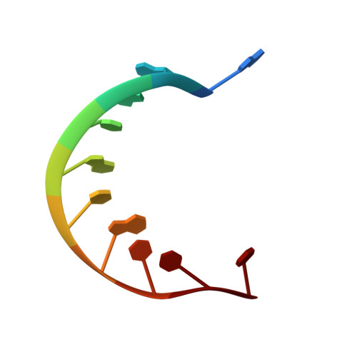
The high resolution crystal structure of the DNA decamer d(AGGCATGCCT).

Nunn, C.M.Neidle, S.

(1996) J Mol Biol 256: 340-351

  • DOI: https://doi.org/10.1006/jmbi.1996.0090
  • Primary Citation of Related Structures:  
    232D

  • PubMed Abstract: 

    The crystal structure of the DNA decamer d(AGGCATGCCT) has been determined to a resolution of 1.3 A and R factor of 13.9%. The structure has a unique conformation with each of the decamer single strands forming base-pairing interactions with two symmetry-related strands. The central eight bases of the decamer form an A-DNA octamer duplex with one symmetry-related strand whilst the terminal 5'-A and T-3' bases are flipped out and away from the octamer helix axis to form base-pairing interactions with a second symmetry-related strand. These A.T base-pairs lie perpendicular to the crystallographic c axis and pack within the unit cell in conjunction with a symmetry-related A.T base-pair displaced by 3.4 A degrees along the c axis. A novel base triplet interaction of the type A*(G.C) is present in the structure with interaction from the major groove side of the terminal 5'-A base to the minor groove of the central A-DNA octamer. This structure reports the first example of cobalt hexammine binding to a right-handed DNA duplex. The crystallographic asymmetric unit contains two cobalt hexammine ligands with one site in the major groove coordinating via hydrogen bonds to the 5'-AGG bases, and the second site located between DNA molecules and interacting with the oxygen atoms of phosphate groups.


  • Organizational Affiliation

    The CRC Biomolecular Structure Unit, The Institute of Cancer Research, Sutton Surrey, UK.


Macromolecules

Find similar nucleic acids by:  Sequence   |   3D Structure  

Entity ID: 1
MoleculeChains LengthOrganismImage
DNA (5'-D(*AP*GP*GP*CP*AP*TP*GP*CP*CP*T)-3')10N/A
Sequence Annotations
Expand
  • Reference Sequence
Small Molecules
Ligands 1 Unique
IDChains Name / Formula / InChI Key2D Diagram3D Interactions
NCO
Query on NCO

Download Ideal Coordinates CCD File 
B [auth A],
C [auth A]
COBALT HEXAMMINE(III)
Co H18 N6
DYLMFCCYOUSRTK-UHFFFAOYSA-N
Experimental Data & Validation

Experimental Data

  • Method: X-RAY DIFFRACTION
  • Resolution: 1.30 Å
  • R-Value Free: 0.216 
  • R-Value Observed: 0.139 
  • Space Group: P 61 2 2
Unit Cell:
Length ( Å )Angle ( ˚ )
a = 32.7α = 90
b = 32.7β = 90
c = 78.7γ = 120
Software Package:
Software NamePurpose
SHELXL-93refinement
DENZOdata reduction

Structure Validation

View Full Validation Report


Entry History 

Deposition Data

Revision History  (Full details and data files)

  • Version 1.0: 1996-03-22
    Type: Initial release
  • Version 1.1: 2008-05-22
    Changes: Version format compliance
  • Version 1.2: 2011-07-13
    Changes: Version format compliance
  • Version 1.3: 2024-02-14
    Changes: Data collection, Database references, Derived calculations