1ZEQ

1.5 A Structure of apo-CusF residues 6-88 from Escherichia coli


Experimental Data Snapshot

  • Method: X-RAY DIFFRACTION
  • Resolution: 1.50 Å
  • R-Value Free: 0.207 
  • R-Value Work: 0.185 
  • R-Value Observed: 0.186 

wwPDB Validation   3D Report Full Report


This is version 1.3 of the entry. See complete history


Literature

A Novel Copper-Binding Fold for the Periplasmic Copper Resistance Protein CusF.

Loftin, I.R.Franke, S.Roberts, S.A.Weichsel, A.Montfort, W.R.Rensing, C.McEvoy, M.M.

(2005) Biochemistry 44: 10533-10540

  • DOI: https://doi.org/10.1021/bi050827b
  • Primary Citation of Related Structures:  
    1ZEQ

  • PubMed Abstract: 

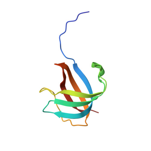
    We have determined the crystal structure of apo-CusF, a periplasmic protein involved in copper and silver resistance in Escherichia coli. The protein forms a five-stranded beta-barrel, classified as an OB-fold, which is a unique topology for a copper-binding protein. NMR chemical shift mapping experiments suggest that Cu(I) is bound by conserved residues H36, M47, and M49 located in beta-strands 2 and 3. These residues are clustered at one end of the beta-barrel, and their side chains are oriented toward the interior of the barrel. Cu(I) can be modeled into the apo-CusF structure with only minimal structural changes using H36, M47, and M49 as ligands. The unique structure and metal binding site of CusF are distinct from those of previously characterized copper-binding proteins.


  • Organizational Affiliation

    Department of Biochemistry and Molecular Biophysics, University of Arizona, Tucson, Arizona 85721, USA.


Macromolecules
Find similar proteins by:  (by identity cutoff)  |  3D Structure
Entity ID: 1
MoleculeChains Sequence LengthOrganismDetailsImage
Cation efflux system protein cusFA [auth X]84Escherichia coliMutation(s): 0 
Gene Names: cusFcusX
UniProt
Find proteins for P77214 (Escherichia coli (strain K12))
Explore P77214 
Go to UniProtKB:  P77214
Entity Groups  
Sequence Clusters30% Identity50% Identity70% Identity90% Identity95% Identity100% Identity
UniProt GroupP77214
Sequence Annotations
Expand
  • Reference Sequence
Experimental Data & Validation

Experimental Data

  • Method: X-RAY DIFFRACTION
  • Resolution: 1.50 Å
  • R-Value Free: 0.207 
  • R-Value Work: 0.185 
  • R-Value Observed: 0.186 
  • Space Group: P 21 21 21
Unit Cell:
Length ( Å )Angle ( ˚ )
a = 39.3α = 90
b = 39.326β = 90
c = 43.693γ = 90
Software Package:
Software NamePurpose
REFMACrefinement
CBASSdata collection
SCALEPACKdata scaling
SOLVEphasing

Structure Validation

View Full Validation Report


Entry History 

Deposition Data

Revision History  (Full details and data files)

  • Version 1.0: 2005-08-02
    Type: Initial release
  • Version 1.1: 2008-04-30
    Changes: Version format compliance
  • Version 1.2: 2011-07-13
    Changes: Version format compliance
  • Version 1.3: 2024-02-14
    Changes: Data collection, Database references