1Y9I

Crystal structure of low temperature requirement C protein from Listeria monocytogenes


Experimental Data Snapshot

  • Method: X-RAY DIFFRACTION
  • Resolution: 1.80 Å
  • R-Value Free: 0.245 
  • R-Value Work: 0.211 

wwPDB Validation   3D Report Full Report


This is version 1.4 of the entry. See complete history


Literature

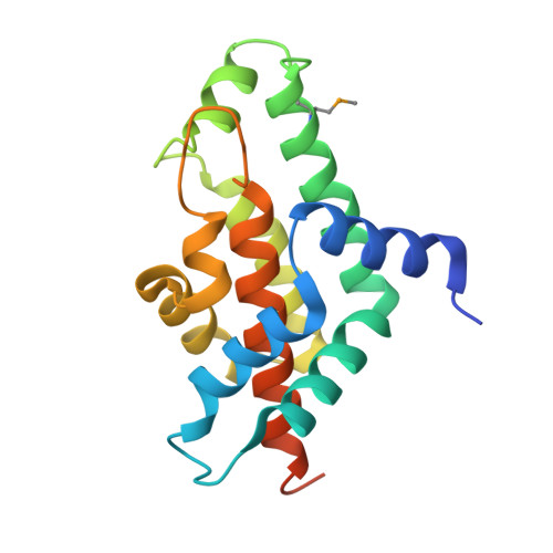
Crystal structure of phosphatidylglycerophosphatase (PGPase), a putative membrane-bound lipid phosphatase, reveals a novel binuclear metal binding site and two "proton wires".

Kumaran, D.Bonanno, J.B.Burley, S.K.Swaminathan, S.

(2006) Proteins 64: 851-862

  • DOI: https://doi.org/10.1002/prot.21039
  • Primary Citation of Related Structures:  
    1Y9I

  • PubMed Abstract: 

    Phosphatidylglycerophosphatase (PGPase), an enzyme involved in lipid metabolism, catalyzes formation of phosphatidylglycerol from phosphatidylglycerophosphate. Phosphatidylglycerol is a multifunctional phospholipid, found in the biological membranes of many organisms. Here, we report the crystal structure of Listeria monocytogenes PGPase at 1.8 A resolution. PGPase, an all-helical molecule, forms a homotetramer. Each protomer contains an independent active site with two metal ions, Ca(2+) and Mg(2+), forming a hetero-binuclear center located in a hydrophilic cavity near the surface of the molecule. The binuclear center, conserved ligands, metal-bound water molecules, and an Asp-His dyad form the active site. The catalytic mechanism of this enzyme is likely to proceed via binuclear metal activated nucleophilic water. The binuclear metal-binding active-site environment of this structure should provide insights into substrate binding and metal-dependent catalysis. A long channel with inter-linked linear water chains, termed "proton wires," is observed at the tetramer interface. Comparison of similar water chain structures in photosynthetic reaction centers (RCs), Cytochrome f, gramicidin, and bacteriorhodopsin, suggests that PGPase may conduct protons via proton wires.


  • Organizational Affiliation

    Biology Department, Brookhaven National Laboratory, Upton, New York 11973, USA.


Macromolecules
Find similar proteins by:  (by identity cutoff)  |  3D Structure
Entity ID: 1
MoleculeChains Sequence LengthOrganismDetailsImage
low temperature requirement C protein
A, B, C, D
178Listeria monocytogenesMutation(s): 1 
UniProt
Find proteins for Q8Y4N7 (Listeria monocytogenes serovar 1/2a (strain ATCC BAA-679 / EGD-e))
Explore Q8Y4N7 
Go to UniProtKB:  Q8Y4N7
Entity Groups  
Sequence Clusters30% Identity50% Identity70% Identity90% Identity95% Identity100% Identity
UniProt GroupQ8Y4N7
Sequence Annotations
Expand
  • Reference Sequence
Small Molecules
Ligands 3 Unique
IDChains Name / Formula / InChI Key2D Diagram3D Interactions
GOL
Query on GOL

Download Ideal Coordinates CCD File 
I [auth B]GLYCEROL
C3 H8 O3
PEDCQBHIVMGVHV-UHFFFAOYSA-N
CA
Query on CA

Download Ideal Coordinates CCD File 
E [auth A],
G [auth B],
J [auth C],
L [auth D]
CALCIUM ION
Ca
BHPQYMZQTOCNFJ-UHFFFAOYSA-N
MG
Query on MG

Download Ideal Coordinates CCD File 
F [auth A],
H [auth B],
K [auth C],
M [auth D]
MAGNESIUM ION
Mg
JLVVSXFLKOJNIY-UHFFFAOYSA-N
Modified Residues  1 Unique
IDChains TypeFormula2D DiagramParent
MSE
Query on MSE
A, B, C, D
L-PEPTIDE LINKINGC5 H11 N O2 SeMET
Experimental Data & Validation

Experimental Data

  • Method: X-RAY DIFFRACTION
  • Resolution: 1.80 Å
  • R-Value Free: 0.245 
  • R-Value Work: 0.211 
  • Space Group: P 1 21 1
Unit Cell:
Length ( Å )Angle ( ˚ )
a = 55.908α = 90
b = 101.069β = 95.4
c = 63.096γ = 90
Software Package:
Software NamePurpose
MAR345data collection
HKL-2000data reduction
SOLVEphasing
SHARPphasing
ARP/wARPmodel building
CNSrefinement
HKL-2000data scaling

Structure Validation

View Full Validation Report


Entry History 

Deposition Data

Revision History  (Full details and data files)

  • Version 1.0: 2004-12-28
    Type: Initial release
  • Version 1.1: 2008-04-30
    Changes: Version format compliance
  • Version 1.2: 2011-07-13
    Changes: Non-polymer description, Version format compliance
  • Version 1.3: 2017-10-11
    Changes: Refinement description
  • Version 1.4: 2021-02-03
    Changes: Database references, Derived calculations, Structure summary