1XEL

UDP-GALACTOSE 4-EPIMERASE FROM ESCHERICHIA COLI


Experimental Data Snapshot

  • Method: X-RAY DIFFRACTION
  • Resolution: 1.80 Å
  • R-Value Observed: 0.137 

wwPDB Validation   3D Report Full Report


This is version 1.4 of the entry. See complete history


Literature

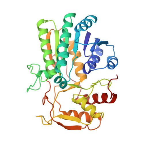
Molecular structure of the NADH/UDP-glucose abortive complex of UDP-galactose 4-epimerase from Escherichia coli: implications for the catalytic mechanism.

Thoden, J.B.Frey, P.A.Holden, H.M.

(1996) Biochemistry 35: 5137-5144

  • DOI: https://doi.org/10.1021/bi9601114
  • Primary Citation of Related Structures:  
    1XEL

  • PubMed Abstract: 

    UDP-galactose 4-epimerase is one of three enzymes in the metabolic pathway that converts galactose into glucose1-phosphate. Specifically this enzyme catalyzes the interconversion of UDP-galactose and UDP-glucose. The molecular structure of the NADH/UDP-glucose abortive complex of the enzyme from Escherichia coli has been determined by X-ray diffraction analysis to a nominal resolution of 1.8 A and refined to an R-factor of 18.2% for all measurement X-ray data. The nicotinamide ring of the dinucleotide adopts the syn conformation in relationship to the ribose. Both the NADH and UDP-glucose are in the proper orientation for a B-side specific transfer from C4 of the sugar to C4 of the dinucleotide. Those residues implicated in glucose binding include Ser 124, tyr 149, Asn 179, Asn199, Arg 231, and Tyr 299. An amino acid sequence alignment of various prokaryotic and eukaryotic epimerases reveals a high degree of conservation with respect to those residues involved in both NADH and substrate binding. The nonstereospecificity displayed by epimerase was originally thought to occur through a simple rotation about the bond between the glycosyl C1 oxygen of the 4-ketose intermediate and the beta-phosphorous of the UDP moiety, thereby allowing the opposite side of the sugar to face the NADH. The present structure reveals that additional rotations about the phosphate backbone of UDP are necessary. Furthermore, the abortive complex model described here suggests that Ser 124 and Tyr 149 are likely to play important roles in the catalytic mechanism of the enzyme.


  • Organizational Affiliation

    Institute for Enzyme Research, Graduate School, University of Wisconsin, Madison 53705, USA.


Macromolecules
Find similar proteins by:  (by identity cutoff)  |  3D Structure
Entity ID: 1
MoleculeChains Sequence LengthOrganismDetailsImage
UDP-GALACTOSE 4-EPIMERASE338Escherichia coliMutation(s): 0 
EC: 5.1.3.2
UniProt
Find proteins for P09147 (Escherichia coli (strain K12))
Explore P09147 
Go to UniProtKB:  P09147
Entity Groups  
Sequence Clusters30% Identity50% Identity70% Identity90% Identity95% Identity100% Identity
UniProt GroupP09147
Sequence Annotations
Expand
  • Reference Sequence
Small Molecules
Ligands 5 Unique
IDChains Name / Formula / InChI Key2D Diagram3D Interactions
NAD
Query on NAD

Download Ideal Coordinates CCD File 
D [auth A]NICOTINAMIDE-ADENINE-DINUCLEOTIDE
C21 H27 N7 O14 P2
BAWFJGJZGIEFAR-NNYOXOHSSA-N
UPG
Query on UPG

Download Ideal Coordinates CCD File 
E [auth A]URIDINE-5'-DIPHOSPHATE-GLUCOSE
C15 H24 N2 O17 P2
HSCJRCZFDFQWRP-JZMIEXBBSA-N
PEG
Query on PEG

Download Ideal Coordinates CCD File 
G [auth A]DI(HYDROXYETHYL)ETHER
C4 H10 O3
MTHSVFCYNBDYFN-UHFFFAOYSA-N
EDO
Query on EDO

Download Ideal Coordinates CCD File 
F [auth A]1,2-ETHANEDIOL
C2 H6 O2
LYCAIKOWRPUZTN-UHFFFAOYSA-N
NA
Query on NA

Download Ideal Coordinates CCD File 
B [auth A],
C [auth A]
SODIUM ION
Na
FKNQFGJONOIPTF-UHFFFAOYSA-N
Experimental Data & Validation

Experimental Data

  • Method: X-RAY DIFFRACTION
  • Resolution: 1.80 Å
  • R-Value Observed: 0.137 
  • Space Group: P 32 2 1
Unit Cell:
Length ( Å )Angle ( ˚ )
a = 83.8α = 90
b = 83.8β = 90
c = 108.4γ = 120
Software Package:
Software NamePurpose
TNTrefinement

Structure Validation

View Full Validation Report


Entry History 

Deposition Data

Revision History  (Full details and data files)

  • Version 1.0: 1997-02-12
    Type: Initial release
  • Version 1.1: 2008-03-03
    Changes: Version format compliance
  • Version 1.2: 2011-07-13
    Changes: Derived calculations, Version format compliance
  • Version 1.3: 2011-11-16
    Changes: Atomic model
  • Version 1.4: 2024-02-14
    Changes: Data collection, Database references, Derived calculations, Other