1XCV

Crystal Structure Of (H79AC102D)Dtxr complexed with Nickel(II)


Experimental Data Snapshot

  • Method: X-RAY DIFFRACTION
  • Resolution: 2.10 Å
  • R-Value Free: 0.300 
  • R-Value Work: 0.279 
  • R-Value Observed: 0.279 

wwPDB Validation   3D Report Full Report


This is version 1.4 of the entry. See complete history


Literature

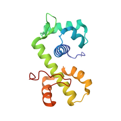
Mechanism of metal ion activation of the diphtheria toxin repressor DtxR.

D'Aquino, J.A.Tetenbaum-Novatt, J.White, A.Berkovitch, F.Ringe, D.

(2005) Proc Natl Acad Sci U S A 102: 18408-18413

  • DOI: https://doi.org/10.1073/pnas.0500908102
  • Primary Citation of Related Structures:  
    1XCV

  • PubMed Abstract: 

    The diphtheria toxin repressor (DtxR) is a metal ion-activated transcriptional regulator that has been linked to the virulence of Corynebacterium diphtheriae. Structure determination has shown that there are two metal ion binding sites per repressor monomer, and site-directed mutagenesis has demonstrated that binding site 2 (primary) is essential for recognition of the target DNA repressor, leaving the role of binding site 1 (ancillary) unclear. Calorimetric techniques have demonstrated that although binding site 1 (ancillary) has high affinity for metal ion with a binding constant of 2 x 10(-7), binding site 2 (primary) is a low-affinity binding site with a binding constant of 6.3 x 10(-4). These two binding sites act in an independent fashion, and their contribution can be easily dissected by traditional mutational analysis. Our results clearly demonstrate that binding site 1 (ancillary) is the first one to be occupied during metal ion activation, playing a critical role in stabilization of the repressor. In addition, structural data obtained for the mutants Ni-DtxR(H79A,C102D), reported here, and the previously reported DtxR(H79A) have allowed us to propose a mechanism of metal activation for DtxR.


  • Organizational Affiliation

    Department of Chemistry, Brandeis University, Waltham, MA 02454, USA.


Macromolecules
Find similar proteins by:  (by identity cutoff)  |  3D Structure
Entity ID: 1
MoleculeChains Sequence LengthOrganismDetailsImage
Diphtheria toxin repressor mutant139Corynebacterium diphtheriaeMutation(s): 2 
Gene Names: dtxR
UniProt
Find proteins for P0DJL7 (Corynebacterium diphtheriae (strain ATCC 700971 / NCTC 13129 / Biotype gravis))
Explore P0DJL7 
Go to UniProtKB:  P0DJL7
Entity Groups  
Sequence Clusters30% Identity50% Identity70% Identity90% Identity95% Identity100% Identity
UniProt GroupP0DJL7
Sequence Annotations
Expand
  • Reference Sequence
Small Molecules
Ligands 1 Unique
IDChains Name / Formula / InChI Key2D Diagram3D Interactions
NI
Query on NI

Download Ideal Coordinates CCD File 
B [auth A]NICKEL (II) ION
Ni
VEQPNABPJHWNSG-UHFFFAOYSA-N
Experimental Data & Validation

Experimental Data

  • Method: X-RAY DIFFRACTION
  • Resolution: 2.10 Å
  • R-Value Free: 0.300 
  • R-Value Work: 0.279 
  • R-Value Observed: 0.279 
  • Space Group: P 31 2 1
Unit Cell:
Length ( Å )Angle ( ˚ )
a = 63.082α = 90
b = 63.082β = 90
c = 105.987γ = 120
Software Package:
Software NamePurpose
CNSrefinement
d*TREKdata reduction
d*TREKdata scaling
EPMRphasing

Structure Validation

View Full Validation Report


Entry History 

Deposition Data

Revision History  (Full details and data files)

  • Version 1.0: 2005-08-16
    Type: Initial release
  • Version 1.1: 2008-04-30
    Changes: Version format compliance
  • Version 1.2: 2011-07-13
    Changes: Derived calculations, Version format compliance
  • Version 1.3: 2021-10-20
    Changes: Database references, Derived calculations
  • Version 1.4: 2023-08-23
    Changes: Data collection, Refinement description