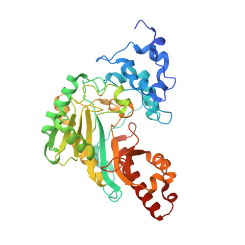
The 2.1 A Structure of Torpedo californica Creatine Kinase Complexed with the ADP-Mg(2+)-NO3(-)-Creatine Transition-State Analogue Complex
Lahiri, S.D., Wang, P.F., Babbitt, P.C., McLeish, M.J., Kenyon, G.L., Allen, K.N.(2002) Biochemistry 41: 13861-13867
- PubMed: 12437342
- DOI: https://doi.org/10.1021/bi026655p
- Primary Citation of Related Structures:
1VRP - PubMed Abstract:
Creatine kinase (CK) catalyzes the reversible conversion of creatine and ATP to phosphocreatine and ADP, thereby helping maintain energy homeostasis in the cell. Here we report the first X-ray structure of CK bound to a transition-state analogue complex (CK-TSAC). Cocrystallization of the enzyme from Torpedo californica (TcCK) with ADP-Mg(2+), nitrate, and creatine yielded a homodimer, one monomer of which was liganded to a TSAC complex while the second monomer was bound to ADP-Mg(2+) alone. The structures of both monomers were determined to 2.1 A resolution. The creatine is located with the guanidino nitrogen cis to the methyl group positioned to perform in-line attack at the gamma-phosphate of ATP-Mg(2+), while the ADP-Mg(2+) is in a conformation similar to that found in the TSAC-bound structure of the homologue arginine kinase (AK). Three ligands to Mg(2+) are contributed by ADP and nitrate and three by ordered water molecules. The most striking difference between the substrate-bound and TSAC-bound structures is the movement of two loops, comprising residues 60-70 and residues 323-332. In the TSAC-bound structure, both loops move into the active site, resulting in the positioning of two hydrophobic residues (one from each loop), Ile69 and Val325, near the methyl group of creatine. This apparently provides a specificity pocket for optimal creatine binding as this interaction is missing in the AK structure. In addition, the active site of the transition-state analogue complex is completely occluded from solvent, unlike the ADP-Mg(2+)-bound monomer and the unliganded structures reported previously.
Organizational Affiliation:
Department of Physiology and Biophysics, Boston University School of Medicine, Boston, Massachusetts 02155, USA.