The crystal structure of microtubule-associated protein light chain 3, a mammalian homologue of Saccharomyces cerevisiae Atg8
Sugawara, K., Suzuki, N.N., Fujioka, Y., Mizushima, N., Ohsumi, Y., Inagaki, F.(2004) Genes Cells 9: 611-618
- PubMed: 15265004
- DOI: https://doi.org/10.1111/j.1356-9597.2004.00750.x
- Primary Citation of Related Structures:
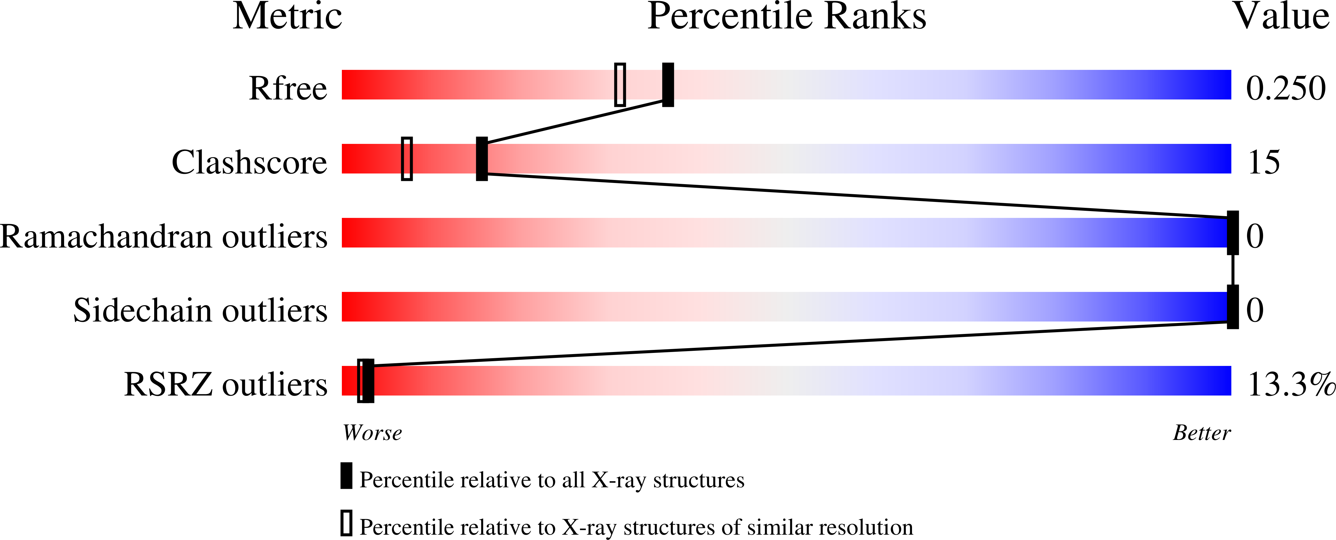
1UGM - PubMed Abstract:
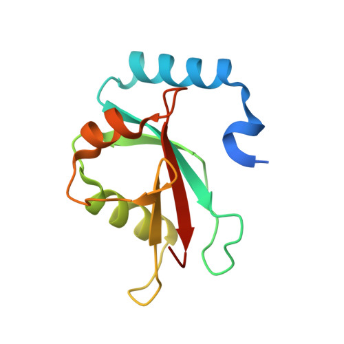
Microtubule-associated protein light chain 3 (LC3), a mammalian homologue of yeast Atg8, plays an essential role in autophagy, which is involved in the bulk degradation of cytoplasmic components by the lysosomal system. Here, we report the crystal structure of LC3 at 2.05 A resolution with an R-factor of 21.8% and a free R-factor of 24.9%. The structure of LC3, which is similar to those of Golgi-associated ATPase enhancer of 16 kDa (GATE-16) and GABAA receptor-associated protein (GABARAP), contains a ubiquitin core with two alpha helices, alpha1 and alpha2, attached at its N-terminus. Some common and distinct features are observed among these proteins, including the conservation of residues required to form an interaction among alpha1, alpha2 and the ubiquitin core. However, the electrostatic potential surfaces of these helices differ, implicating particular roles to select specific binding partners. Hydrophobic patches on the ubiquitin core of LC3, GABARAP and GATE-16 are well conserved and are similar to the E1 binding surface of ubiquitin and NEDD8. Therefore, we propose that the hydrophobic patch is a binding surface for mammalian Atg7 similar to a ubiquitin-like conjugation system. We also propose the functional implications of the ubiquitin fold as a recognition module of target proteins.
Organizational Affiliation:
Department of Structural Biology, Graduate School of Pharmaceutical Sciences, Hokkaido University, Kita-12, Nishi-6, Kita-ku, Sapporo 060-0812, Japan.