1SQC

SQUALENE-HOPENE-CYCLASE FROM ALICYCLOBACILLUS ACIDOCALDARIUS


Experimental Data Snapshot

  • Method: X-RAY DIFFRACTION
  • Resolution: 2.85 Å
  • R-Value Free: 0.243 
  • R-Value Work: 0.167 
  • R-Value Observed: 0.167 

wwPDB Validation   3D Report Full Report


Ligand Structure Quality Assessment 


This is version 1.3 of the entry. See complete history


Literature

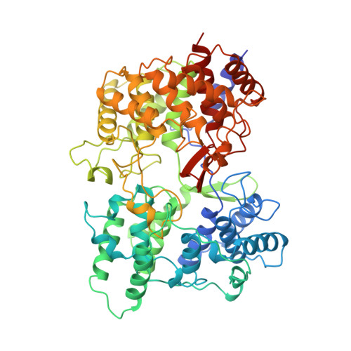
Structure and function of a squalene cyclase.

Wendt, K.U.Poralla, K.Schulz, G.E.

(1997) Science 277: 1811-1815

  • DOI: https://doi.org/10.1126/science.277.5333.1811
  • Primary Citation of Related Structures:  
    1SQC

  • PubMed Abstract: 

    The crystal structure of squalene-hopene cyclase from Alicyclobacillus acidocaldarius was determined at 2.9 angstrom resolution. The mechanism and sequence of this cyclase are closely related to those of 2,3-oxidosqualene cyclases that catalyze the cyclization step in cholesterol biosynthesis. The structure reveals a membrane protein with membrane-binding characteristics similar to those of prostaglandin-H2 synthase, the only other reported protein of this type. The active site of the enzyme is located in a large central cavity that is of suitable size to bind squalene in its required conformation and that is lined by aromatic residues. The structure supports a mechanism in which the acid starting the reaction by protonating a carbon-carbon double bond is an aspartate that is coupled to a histidine. Numerous surface alpha helices are connected by characteristic QW-motifs (Q is glutamine and W is tryptophan) that tighten the protein structure, possibly for absorbing the reaction energy without structural damage.


  • Organizational Affiliation

    Institut für Organische Chemie und Biochemie, Albertstrasse 21, D-79104 Freiburg im Breisgau, Germany.


Macromolecules
Find similar proteins by:  (by identity cutoff)  |  3D Structure
Entity ID: 1
MoleculeChains Sequence LengthOrganismDetailsImage
SQUALENE-HOPENE CYCLASE631Alicyclobacillus acidocaldariusMutation(s): 0 
EC: 5.4.99
UniProt
Find proteins for P33247 (Alicyclobacillus acidocaldarius subsp. acidocaldarius (strain ATCC 27009 / DSM 446 / BCRC 14685 / JCM 5260 / KCTC 1825 / NBRC 15652 / NCIMB 11725 / NRRL B-14509 / 104-IA))
Explore P33247 
Go to UniProtKB:  P33247
Entity Groups  
Sequence Clusters30% Identity50% Identity70% Identity90% Identity95% Identity100% Identity
UniProt GroupP33247
Sequence Annotations
Expand
  • Reference Sequence
Small Molecules
Ligands 1 Unique
IDChains Name / Formula / InChI Key2D Diagram3D Interactions
LDA
Query on LDA

Download Ideal Coordinates CCD File 
B [auth A]LAURYL DIMETHYLAMINE-N-OXIDE
C14 H31 N O
SYELZBGXAIXKHU-UHFFFAOYSA-N
Binding Affinity Annotations 
IDSourceBinding Affinity
LDA PDBBind:  1SQC Ki: 140 (nM) from 1 assay(s)
Experimental Data & Validation

Experimental Data

  • Method: X-RAY DIFFRACTION
  • Resolution: 2.85 Å
  • R-Value Free: 0.243 
  • R-Value Work: 0.167 
  • R-Value Observed: 0.167 
  • Space Group: P 3 2 1
Unit Cell:
Length ( Å )Angle ( ˚ )
a = 140.7α = 90
b = 140.7β = 90
c = 81.9γ = 120
Software Package:
Software NamePurpose
X-PLORmodel building
X-PLORrefinement
XDSdata reduction
XSCALEdata scaling
CCP4data scaling
X-PLORphasing

Structure Validation

View Full Validation Report


Ligand Structure Quality Assessment 


Entry History 

Deposition Data

Revision History  (Full details and data files)

  • Version 1.0: 1997-12-17
    Type: Initial release
  • Version 1.1: 2008-03-24
    Changes: Version format compliance
  • Version 1.2: 2011-07-13
    Changes: Version format compliance
  • Version 1.3: 2024-02-14
    Changes: Data collection, Database references, Derived calculations, Other