1SF2

Structure of E. coli gamma-aminobutyrate aminotransferase


Experimental Data Snapshot

  • Method: X-RAY DIFFRACTION
  • Resolution: 2.40 Å
  • R-Value Free: 0.211 
  • R-Value Work: 0.165 
  • R-Value Observed: 0.167 

wwPDB Validation   3D Report Full Report


This is version 1.4 of the entry. See complete history


Literature

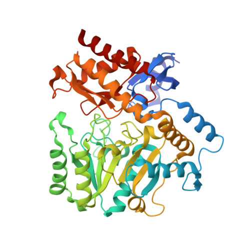
Crystal structures of unbound and aminooxyacetate-bound Escherichia coli gamma-aminobutyrate aminotransferase.

Liu, W.Peterson, P.E.Carter, R.J.Zhou, X.Langston, J.A.Fisher, A.J.Toney, M.D.

(2004) Biochemistry 43: 10896-10905

  • DOI: https://doi.org/10.1021/bi049218e
  • Primary Citation of Related Structures:  
    1SF2, 1SFF

  • PubMed Abstract: 

    The X-ray crystal structures of Escherichia coli gamma-aminobutyrate aminotransferase unbound and bound to the inhibitor aminooxyacetate are reported. The enzyme crystallizes from ammonium sulfate solutions in the P3(2)21 space group with a tetramer in the asymmetric unit. Diffraction data were collected to 2.4 A resolution for the unliganded enzyme and 1.9 A resolution for the aminooxyacetate complex. The overall structure of the enzyme is similar to those of other aminotransferase subgroup II enzymes. The ability of gamma-aminobutyrate aminotransferase to act on primary amine substrates (gamma-aminobutyrate) in the first half-reaction and alpha-amino acids in the second is proposed to be enabled by the presence of Glu211, whose side chain carboxylate alternates between interactions with Arg398 in the primary amine half-reaction and an alternative binding site in the alpha-amino acid half-reaction, in which Arg398 binds the substrate alpha-carboxylate. The specificity for a carboxylate group on the substrate side chain is due primarily to the presence of Arg141, but also requires substantial local main chain rearrangements relative to the structurally homologous enzyme dialkylglycine decarboxylase, which is specific for small alkyl side chains. No iron-sulfur cluster is found in the bacterial enzyme as was found in the pig enzyme [Storici, P., De Biase, D., Bossa, F., Bruno, S., Mozzarelli, A., Peneff, C., Silverman, R. B., and Schirmer, T. (2004) J. Biol. Chem. 279, 363-73.]. The binding of aminooxyacetate causes remarkably small changes in the active site structure, and no large domain movements are observed. Active site structure comparisons with pig gamma-aminobutyrate aminotransferase and dialkylglycine decarboxylase are discussed.


  • Organizational Affiliation

    Department of Chemistry, University of California, Davis, California 95616, USA.


Macromolecules
Find similar proteins by:  (by identity cutoff)  |  3D Structure
Entity ID: 1
MoleculeChains Sequence LengthOrganismDetailsImage
4-aminobutyrate aminotransferase
A, B, C, D
426Escherichia coliMutation(s): 0 
Gene Names: GABTB2662
EC: 2.6.1.19
UniProt
Find proteins for P22256 (Escherichia coli (strain K12))
Explore P22256 
Go to UniProtKB:  P22256
Entity Groups  
Sequence Clusters30% Identity50% Identity70% Identity90% Identity95% Identity100% Identity
UniProt GroupP22256
Sequence Annotations
Expand
  • Reference Sequence
Small Molecules
Ligands 3 Unique
IDChains Name / Formula / InChI Key2D Diagram3D Interactions
PLP
Query on PLP

Download Ideal Coordinates CCD File 
BA [auth D],
G [auth A],
N [auth B],
S [auth C]
PYRIDOXAL-5'-PHOSPHATE
C8 H10 N O6 P
NGVDGCNFYWLIFO-UHFFFAOYSA-N
SO4
Query on SO4

Download Ideal Coordinates CCD File 
AA [auth D]
E [auth A]
F [auth A]
K [auth B]
L [auth B]
AA [auth D],
E [auth A],
F [auth A],
K [auth B],
L [auth B],
M [auth B],
Q [auth C],
R [auth C],
X [auth D],
Y [auth D],
Z [auth D]
SULFATE ION
O4 S
QAOWNCQODCNURD-UHFFFAOYSA-L
EDO
Query on EDO

Download Ideal Coordinates CCD File 
CA [auth D]
DA [auth D]
EA [auth D]
FA [auth D]
H [auth A]
CA [auth D],
DA [auth D],
EA [auth D],
FA [auth D],
H [auth A],
I [auth A],
J [auth A],
O [auth B],
P [auth B],
T [auth C],
U [auth C],
V [auth C],
W [auth C]
1,2-ETHANEDIOL
C2 H6 O2
LYCAIKOWRPUZTN-UHFFFAOYSA-N
Experimental Data & Validation

Experimental Data

  • Method: X-RAY DIFFRACTION
  • Resolution: 2.40 Å
  • R-Value Free: 0.211 
  • R-Value Work: 0.165 
  • R-Value Observed: 0.167 
  • Space Group: P 32 2 1
Unit Cell:
Length ( Å )Angle ( ˚ )
a = 108.09α = 90
b = 108.09β = 90
c = 301.25γ = 120
Software Package:
Software NamePurpose
SCALAdata scaling
CNSrefinement
CCP4data scaling
CNSphasing

Structure Validation

View Full Validation Report


Entry History 

Deposition Data

Revision History  (Full details and data files)

  • Version 1.0: 2004-09-14
    Type: Initial release
  • Version 1.1: 2008-04-29
    Changes: Version format compliance
  • Version 1.2: 2011-07-13
    Changes: Non-polymer description, Version format compliance
  • Version 1.3: 2017-10-11
    Changes: Refinement description
  • Version 1.4: 2018-01-31
    Changes: Experimental preparation