1SDX

Crystal structure of the zinc saturated C-terminal half of bovine lactoferrin at 2.0 A resolution reveals two additional zinc binding sites


Experimental Data Snapshot

  • Method: X-RAY DIFFRACTION
  • Resolution: 2.06 Å
  • R-Value Free: 0.210 
  • R-Value Work: 0.192 
  • R-Value Observed: 0.192 

wwPDB Validation   3D Report Full Report


This is version 2.1 of the entry. See complete history


Literature

Structure of the zinc-saturated C-terminal lobe of bovine lactoferrin at 2.0 A resolution.

Jabeen, T.Sharma, S.Singh, N.Bhushan, A.Singh, T.P.

(2005) Acta Crystallogr D Biol Crystallogr 61: 1107-1115

  • DOI: https://doi.org/10.1107/S0907444905016069
  • Primary Citation of Related Structures:  
    1SDX

  • PubMed Abstract: 

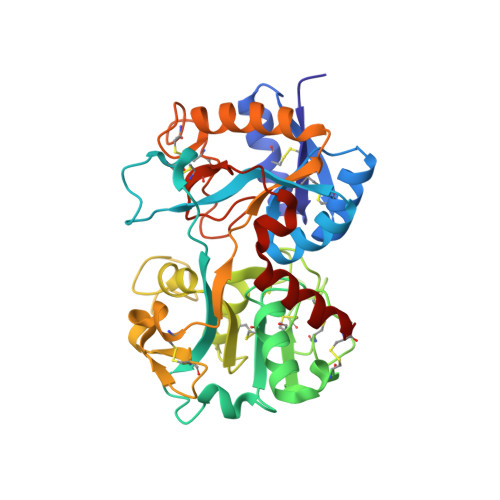
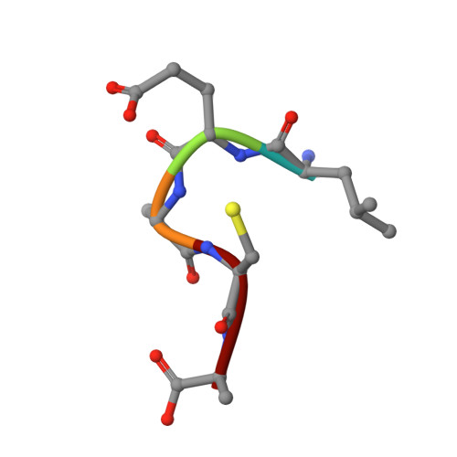
    The crystal structure of the zinc-saturated C-terminal lobe of bovine lactoferrin has been determined at 2.0 A resolution using crystals stabilized at pH 3.8. This is the first metal-saturated structure of any functional lactoferrin at such a low pH. Purified samples of proteolytically generated zinc-saturated C-terminal lobe were crystallized from 0.1 M MES buffer pH 6.5 containing 25%(v/v) polyethyleneglycol monomethyl ether 550 and 0.1 M zinc sulfate heptahydrate. The crystals were transferred to 25 mM ammonium acetate buffer containing 25%(v/v) polyethyleneglycol monomethyl ether 550 and the pH was gradually changed from 6.5 to 3.8. The X-ray intensity data were collected with a 345 mm imaging-plate scanner mounted on an RU-300 rotating-anode X-ray generator using crystals soaked in the buffer at pH 3.8. The structure was determined with the molecular-replacement method using the coordinates of the monoferric C-terminal lobe of bovine lactoferrin as a search model and was refined to an R factor of 0.192 for all data to 2.0 A resolution. The final model comprises 2593 protein atoms (residues 342-676 and 681-685), 138 carbohydrate atoms (from 11 monosaccharide units in three glycan chains), three Zn2+ ions, one CO3(2-) ion, one SO(4)2- ions and 227 water molecules. The overall folding of the present structure is essentially similar to that of the monoferric C-terminal lobe of bovine lactoferrin, although it contains Zn2+ in place of Fe3+ in the metal-binding cleft as well as two additional Zn2+ ions on the surface of the C-terminal lobe. The Zn2+ ion in the cleft remains bound to the lobe with octahedral coordination. The bidentate carbonate ion is stabilized by a network of hydrogen bonds to Ala465, Gly466, Thr459 and Arg463. The other two zinc ions also form sixfold coordinations involving symmetry-related protein and water molecules. The number of monosaccharide residues from the three glycan chains of the C-terminal lobe was 11, which is the largest number observed to date. The structure shows that the C-terminal lobe of lactoferrin is capable of sequestering a Zn2+ ion at a pH of 3.8. This implies that the zinc ions can be sequestered over a wide pH range. The glycan chain attached to Asn545 may also have some influence on iron release from the C-terminal lobe.


  • Organizational Affiliation

    Department of Biophysics, All India Institute of Medical Sciences, New Delhi 110 029, India.


Macromolecules
Find similar proteins by:  (by identity cutoff)  |  3D Structure
Entity ID: 1
MoleculeChains Sequence LengthOrganismDetailsImage
Lactotransferrin335Bos taurusMutation(s): 0 
UniProt
Find proteins for P24627 (Bos taurus)
Explore P24627 
Go to UniProtKB:  P24627
Entity Groups  
Sequence Clusters30% Identity50% Identity70% Identity90% Identity95% Identity100% Identity
UniProt GroupP24627
Sequence Annotations
Expand
  • Reference Sequence

Find similar proteins by:  Sequence   |   3D Structure  

Entity ID: 2
MoleculeChains Sequence LengthOrganismDetailsImage
LactotransferrinB [auth E]5Bos taurusMutation(s): 0 
UniProt
Find proteins for P24627 (Bos taurus)
Explore P24627 
Go to UniProtKB:  P24627
Entity Groups  
UniProt GroupP24627
Sequence Annotations
Expand
  • Reference Sequence
Oligosaccharides

Help

Entity ID: 3
MoleculeChains Length2D Diagram Glycosylation3D Interactions
2-acetamido-2-deoxy-alpha-D-glucopyranose-(1-4)-2-acetamido-2-deoxy-beta-D-glucopyranoseC [auth B]2N-Glycosylation
Glycosylation Resources
GlyTouCan:  G07375KG
GlyCosmos:  G07375KG
GlyGen:  G07375KG
Entity ID: 4
MoleculeChains Length2D Diagram Glycosylation3D Interactions
alpha-D-mannopyranose-(1-3)-[alpha-D-mannopyranose-(1-4)]alpha-D-mannopyranose-(1-4)-beta-D-mannopyranose-(1-4)-2-acetamido-2-deoxy-alpha-D-glucopyranose-(1-4)-2-acetamido-2-deoxy-beta-D-glucopyranoseD [auth C]6N-Glycosylation
Glycosylation Resources
GlyTouCan:  G86938CX
GlyCosmos:  G86938CX
GlyGen:  G86938CX
Entity ID: 5
MoleculeChains Length2D Diagram Glycosylation3D Interactions
alpha-D-mannopyranose-(1-4)-2-acetamido-2-deoxy-beta-D-glucopyranose-(1-4)-2-acetamido-2-deoxy-beta-D-glucopyranoseE [auth D]3N-Glycosylation
Glycosylation Resources
GlyTouCan:  G62182OO
GlyCosmos:  G62182OO
GlyGen:  G62182OO
Experimental Data & Validation

Experimental Data

  • Method: X-RAY DIFFRACTION
  • Resolution: 2.06 Å
  • R-Value Free: 0.210 
  • R-Value Work: 0.192 
  • R-Value Observed: 0.192 
  • Space Group: P 1 21 1
Unit Cell:
Length ( Å )Angle ( ˚ )
a = 63.21α = 90
b = 50.57β = 107.64
c = 66.17γ = 90
Software Package:
Software NamePurpose
CNSrefinement
MAR345data collection
SCALEPACKdata scaling
AMoREphasing

Structure Validation

View Full Validation Report


Entry History 

Deposition Data

Revision History  (Full details and data files)

  • Version 1.0: 2004-03-02
    Type: Initial release
  • Version 1.1: 2008-04-29
    Changes: Version format compliance
  • Version 1.2: 2011-07-13
    Changes: Non-polymer description, Version format compliance
  • Version 1.3: 2017-10-11
    Changes: Refinement description
  • Version 2.0: 2020-07-29
    Type: Remediation
    Reason: Carbohydrate remediation
    Changes: Advisory, Atomic model, Data collection, Derived calculations, Structure summary
  • Version 2.1: 2023-10-25
    Changes: Data collection, Database references, Refinement description, Structure summary