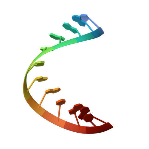
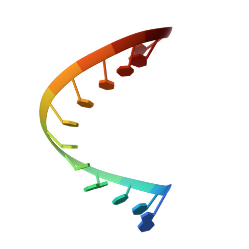
Crystal structure of an RNA dodecamer containing the Escherichia coli Shine-Dalgarno sequence.
Schindelin, H., Zhang, M., Bald, R., Furste, J.P., Erdmann, V.A., Heinemann, U.(1995) J Mol Biol 249: 595-603
- PubMed: 7540215
- DOI: https://doi.org/10.1006/jmbi.1995.0321
- Primary Citation of Related Structures:
1SDR - PubMed Abstract:
The synthetic dodecameric RNA fragment rUAAGGAGGUGAU resembles a region upstream of the initiation site in prokaryotic mRNAs whereas the pyrimidine-rich complementary strand is identical to the last 12 nucleotides of Escherichia coli 16 S rRNA. The complex thus serves as a model for the Shine-Dalgarno interaction which is required for proper initiation of translation. The crystal structure of rUAAGGAGGUGUA.rAUCACCUCCUUA has been determined at 2.6 A resolution and refined against 2957 1 sigma(F) structure amplitudes to an R-value of 0.195. The unit cell of the triclinic crystals contains two double-stranded RNA molecules. The conformation of the two duplexes is similar, with a root-mean-square deviation of 0.683 A between equivalent atoms, and resembles calf thymus A-DNA as determined by X-ray fiber diffraction methods. Both molecules from continuous helices that penetrate the entire crystal, but the dinucleotide step in between dodecameric duplexes has an unusual geometry with a negative twist angle. The long helices cross over each other in a characteristic manner by inserting the backbone of one molecule into the minor groove of another. These contacts are stabilized by several direct intermolecular hydrogen bonds most of which are mediated by 2'-hydroxyl groups of the ribose sugars suggesting a general mode for the interaction between RNA molecules which is different from DNA-DNA interactions.
Organizational Affiliation:
Forschungsgruppe Kristallographie Max-Delbrück-Centrum für Molekulare Medizin, Berlin, FRG.